File:Zhai et al. 2021 f1.jpg

Size of this preview: 735 × 599 pixels. Other resolutions: 294 × 240 pixels | 589 × 480 pixels | 942 × 768 pixels | 1,256 × 1,024 pixels | 1,280 × 1,044 pixels.
Original file (1,280 × 1,044 pixels, file size: 266 KB, MIME type: image/jpeg)
Captions
Captions
Micro-CT images of Chuandianella ovata
Summary
edit
| Description |
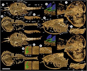
English: Figure 1. Micro-CT images of Chuandianella ovata. (A,C-F) YKLP 16218. (A) Ventral view. Scale bar = 5.0 mm. (C) Endopod of 6th appendage showing endites. Scale bar = 1.6 mm. (D) Ventral view (white rectangle in A), showing details of second appendage (a2, green). Scale bar = 2.0 mm. (E) Same as D but with a2 not coloured. Scale bar = 2.0 mm. (F) Close-up view of 2nd appendages (rotated by 90 degrees with anterior to upper position), with podomeres numbered. Scale bar = 1.4 mm. (B,G) YKLP 16216. (B) Taphonomically deformed specimen, showing ventral view of the carapace and dorsal view of the soft parts (cf. Supplementary Figure 1). Scale bar = 3.8 mm. (G) Dorsal view of part of right side of the body, showing exopods (blue) and endopods (green). Scale bar = 1.4 mm. (H) YKLP 16238, left lateral view, showing endopods and exopods of trunk appendages. Scale bar = 4.9 mm. (I,J) YKLP 16239. (I) Ventral view, showing circa 20 eggs within the left valve. Scale bar = 4.3 mm. (J) Endopods of 3rd and 5th (?) appendages showing long blade-like endites each with a terminal seta. Proximally in this image two endopods overlap, giving the false impression of setae along the lateral margins of the endites. Scale bar = 3.4 mm. All panels are stereo-pairs. Abbreviations: a1, antennule; a2, second appendage; a3-a12, biramous appendages; ab1-4, abdominal segments 1 to 4; an, anus; asc, anterior sclerite; cs, caudal structure; eg, egg; en, endopod; es, eye stalk; et, endite; ex, exopod; ey, stalked eyes; lv, left valve; rv, right valve; se, seta; tp, tailpiece; tr, trunk. Italics indicate a right-side appendage. |
| Date | |
| Source |
An exceptionally preserved euarthropod with unique feather-like appendages from the Chengjiang biota Dayou Zhai, Mark Williams, David J. Siveter, Derek J. Siveter, Thomas H.P. Harvey, Robert S. Sansom, Huijuan Mai, Runqing Zhou, Xianguang Hou bioRxiv 2021.01.22.427827; doi: https://doi.org/10.1101/2021.01.22.427827 This article is a preprint and has not been certified by peer review |
| Author | Dayou Zhai, Mark Williams, David J. Siveter, Derek J. Siveter, Thomas H.P. Harvey, Robert S. Sansom, Huijuan Mai, Runqing Zhou, Xianguang Hou |
Licensing
edit
This file is licensed under the Creative Commons Attribution 4.0 International license.
- You are free:
- to share – to copy, distribute and transmit the work
- to remix – to adapt the work
- Under the following conditions:
- attribution – You must give appropriate credit, provide a link to the license, and indicate if changes were made. You may do so in any reasonable manner, but not in any way that suggests the licensor endorses you or your use.
File history
Click on a date/time to view the file as it appeared at that time.
| Date/Time | Thumbnail | Dimensions | User | Comment | |
|---|---|---|---|---|---|
| current | 11:26, 20 June 2024 | | 1,280 × 1,044 (266 KB) | Qohelet12 (talk | contribs) | Uploaded a work by Dayou Zhai, Mark Williams, David J. Siveter, Derek J. Siveter, Thomas H.P. Harvey, Robert S. Sansom, Huijuan Mai, Runqing Zhou, Xianguang Hou from An exceptionally preserved euarthropod with unique feather-like appendages from the Chengjiang biota Dayou Zhai, Mark Williams, David J. Siveter, Derek J. Siveter, Thomas H.P. Harvey, Robert S. Sansom, Huijuan Mai, Runqing Zhou, Xianguang Hou bioRxiv 2021.01.22.427827; doi: https://doi.org/10.1101/2021.01.22.427827 This article... |
You cannot overwrite this file.
File usage on Commons
There are no pages that use this file.