Simulated X-ray phase contrast radiographs
-
 Fig. 1: Phantom object for X-ray phase contrast simulations. Red: fully absorbing disks. Blue: phase-shifting disks with zero absorption. Disk diameter: 5 µm. The whole image corresponds to an area of 50×50 µm2.
 -
 Fig. 2a: Simulated radiograph for illumination of the phantom from Fig. 1 with a plane monochromatic coherent wave of 12.4 keV photon energy, for a detector placed at a distance D=0.1 mm behind the object.
 -
 Fig. 2b: Same as Fig. 2a, but for D=1 mm.
 -
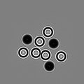 Fig. 2c: Same as Fig. 2a, but for D=10 mm.
 -
 Fig. 2d: Same as Fig. 2a, but for D=100 mm.
 -
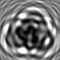 Fig. 2e: Same as Fig. 2a, but for D=1000 mm.


References and additional info edit
The simulation of the wavefront propagation was performed with the XWFP propagation code (T. Weitkamp, Proceedings SPIE 5536 (2004) 181-189), using a Fresnel propagation kernel in paraxial approximation.