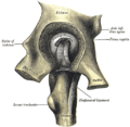
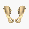
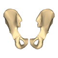
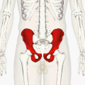
Category:Human hip bone
Subcategories
This category has the following 16 subcategories, out of 16 total.
- SVG human hip bone (2 F)
- Videos of human hip bone (2 F)
3
- 3D data of human hip bone (3 F)
A
- Animations of human hip bone (8 F)
G
I
O
- Obturator foramen (53 F)
P
- Photographs of human hip bone (19 F)
Media in category "Human hip bone"
The following 135 files are in this category, out of 135 total.
-
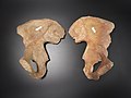
 808 Hip Bone.jpg 974 × 700; 317 KB
-
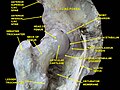
 Atlas and text-book of topographic and applied anatomy (1905) (14803418413).jpg 1,772 × 2,304; 520 KB
-
 Beckenmodell geändert.jpg 359 × 289; 16 KB
-
 BodyParts3D Hip bone.stl 5,120 × 2,880; 230 KB
-
 Braus 1921 225.png 1,758 × 858; 4.32 MB
-
 Braus 1921 227.png 1,038 × 648; 1.93 MB
-
 Braus 1921 228.png 1,422 × 1,254; 5.11 MB
-
 Coxal bone (ilium, ischium and pubis).jpg 960 × 720; 87 KB
-
 Coxal bone - angles.jpg 960 × 720; 109 KB
-
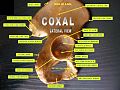
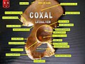
 Coxal bone - lateral view.jpg 960 × 720; 118 KB
-
 Coxal bone - medial view.jpg 960 × 720; 110 KB
-
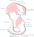
 Coxal bone - muscular insertions.jpg 960 × 720; 94 KB
-
 Coxal bone.jpg 960 × 720; 76 KB
-
 Cunningham’s Text-book of Anatomy (1914) - Fig 230.png 1,428 × 1,584; 2.31 MB
-
 Cunningham’s Text-book of Anatomy (1914) - Fig 232.png 1,668 × 1,274; 1.47 MB
-
 Cunningham’s Text-book of Anatomy (1914) - Fig 233.png 1,406 × 1,176; 1.42 MB
-
 Cunningham’s Text-book of Anatomy (1914) - Fig 369.png 1,872 × 1,587; 1.84 MB
-
 Dixon's Manual of human osteology (1912) - Fig 055.png 1,530 × 1,686; 1.38 MB
-
 Dixon's Manual of human osteology (1912) - Fig 056.png 1,470 × 1,902; 2.13 MB
-
 Dixon's Manual of human osteology (1912) - Fig 057.png 1,536 × 1,875; 1.78 MB
-
 Dixon's Manual of human osteology (1912) - Fig 058.png 1,230 × 1,890; 1.38 MB
-
 Dixon's Manual of human osteology (1912) - Fig 059.png 1,563 × 1,989; 1.57 MB
-
 Dixon's Manual of human osteology (1912) - Fig 060.png 1,464 × 1,624; 1.09 MB
-
 Etching of innominate bone Wellcome V0008824.jpg 2,457 × 3,518; 4.19 MB
-
 Exsection of hip-joint.jpg 1,528 × 1,245; 634 KB
-
 Gauche.jpg 1,552 × 1,055; 41 KB
-
 Genga 05.jpg 1,200 × 1,631; 127 KB
-
 Gerrish's Text-book of Anatomy (1902) - Fig. 177.png 1,452 × 1,443; 1.08 MB
-
 Gerrish's Text-book of Anatomy (1902) - Fig. 178.png 1,407 × 1,332; 957 KB
-
 Gerrish's Text-book of Anatomy (1902) - Fig. 179.png 1,458 × 1,396; 1.15 MB
-
 Gerrish's Text-book of Anatomy (1902) - Fig. 180.png 1,158 × 1,350; 713 KB
-
 Gerrish's Text-book of Anatomy (1902) - Fig. 181.png 776 × 960; 561 KB
-
 GirlSkeletonBodyFront.gif 1,410 × 3,073; 429 KB
-
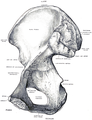
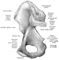
 Gray1244.png 470 × 450; 78 KB
-
 Gray235 he.png 775 × 911; 131 KB
-
 Gray235-ar.png 793 × 911; 533 KB
-
 Gray235.png 793 × 911; 122 KB
-
 Gray236-ar.png 485 × 700; 209 KB
-
 Gray236.png 485 × 700; 49 KB
-
 Gray237-ar.png 430 × 500; 175 KB
-
 Gray237.png 459 × 533; 33 KB
-
 Gray321-ar.png 600 × 458; 220 KB
-
 Gray321.png 600 × 458; 50 KB
-
 Gray321mod.png 1,070 × 908; 234 KB
-
 Gray341 zh.png 512 × 500; 157 KB
-
 Gray341-ar.png 516 × 500; 275 KB
-
 Gray341.png 512 × 500; 47 KB
-
 Gray343.png 444 × 500; 48 KB
-
 Gray430.png 354 × 1,229; 102 KB
-
 Hip bone - close-up - animation (left hip bone).gif 320 × 320; 838 KB
-
 Hip bone - close-up - animation (right hip bone).gif 320 × 320; 787 KB
-
 Hip bone - close-up - animation.gif 320 × 320; 790 KB
-
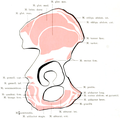
 Hip bone - close-up - anterior view.png 900 × 900; 175 KB
-
 Hip bone - close-up - inferior view animation.gif 320 × 320; 935 KB
-
 Hip bone - close-up - inferior view.png 900 × 900; 149 KB
-
 Hip bone - close-up - lateral view (right hip bone).png 900 × 900; 166 KB
-
 Hip bone - close-up - medial view (left hip bone).png 900 × 900; 154 KB
-
 Hip bone - close-up - medial view (right hip bone).png 900 × 900; 158 KB
-
 Hip bone - close-up - posterior view.png 900 × 900; 171 KB
-
 Hip bone - close-up - superior view.png 900 × 900; 138 KB
-
 Hip bone animation.gif 320 × 320; 1.3 MB
-
 Hip bone animation2.gif 320 × 320; 1.96 MB
-
 Hip bone animation3.gif 320 × 320; 1.08 MB
-
 Hip bone animation4.gif 320 × 320; 1.83 MB
-
 Hip bone anterior high-res.jpg 6,000 × 6,000; 4 MB
-
 Hip bone anterior.png 900 × 900; 358 KB
-
 Hip bone anterior2.png 900 × 900; 416 KB
-
 Hip bone anterior3.png 900 × 900; 360 KB
-
 Hip bone lateral.png 900 × 900; 212 KB
-
 Hip bone lateral2.png 900 × 900; 259 KB
-
 Hip bone lateral3.png 900 × 900; 191 KB
-
 Hip bone posterior.png 900 × 900; 356 KB
-
 Hip bone posterior2.png 900 × 900; 447 KB
-
 Hip.jpg 330 × 310; 9 KB
-
 HK TST Science Museum Bones exhibit 23 人類 skeletons.JPG 2,448 × 3,264; 1.61 MB
-
 Holden's human osteology (1899) - Fig24.png 1,100 × 720; 358 KB
-
 Holden's human osteology (1899) - Plt30 Fig01.png 982 × 1,260; 627 KB
-
 Holden's human osteology (1899) - Plt30 Fig02.png 1,134 × 1,202; 828 KB
-
 Hueter.jpg 257 × 124; 27 KB
-
 Human hip bone texture.jpg 3,872 × 2,592; 5.09 MB
-
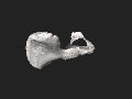
 Human hip bone.stl 5,120 × 2,880; 9.73 MB
-
 Human pelvis.stl 5,120 × 2,880; 20.09 MB
-
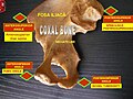
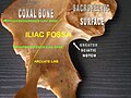 Iliac fossa 2.jpg 960 × 720; 95 KB
-
 Iliac fossa.jpg 960 × 720; 85 KB
-
 Illu pelvic girdle RO mod.jpg 453 × 327; 39 KB
-
 Illu pelvic girdle RO.jpg 350 × 231; 17 KB
-
 Iv3d-torso.png 640 × 616; 615 KB
-
 Laced White Lingerie.png 4,200 × 3,000; 8.62 MB
-
 Leonardo Skeleton 1511.jpg 475 × 594; 96 KB
-
 Merkel's Human Anatomy (1913) - Vol 3 - Fig 119.png 1,500 × 1,476; 723 KB
-
 Merkel's Human Anatomy (1913) - Vol 3 - Fig 120.png 1,326 × 1,449; 858 KB
-
 Morris' human anatomy (1898) - Fig 151.png 1,720 × 1,956; 2.18 MB
-
 Morris' human anatomy (1898) - Fig 152.png 1,695 × 1,926; 2.52 MB
-
 Morris' human anatomy (1933) - Fig 253.png 2,144 × 2,312; 4.29 MB
-
 Morris' human anatomy (1933) - Fig 254.png 2,080 × 2,304; 3.8 MB
-
 Os coxae.stl 5,120 × 2,880; 7.45 MB
-
 Os coxal face externe.png 935 × 911; 488 KB
-
 Os coxal face interne.png 557 × 599; 273 KB
-
 Os coxal face pelvienne.png 415 × 599; 228 KB
-
 Os coxal1 insertions musculairese.png 935 × 911; 473 KB
-
Pelvic Bones Walk-thru.webm 11 min 33 s, 640 × 480; 15.57 MB
-
Pelvic Girdle Anatomy by Jason Christian.webm 1 min 50 s, 1,280 × 720; 16.81 MB
-
 Pelvis - os coxae (dex, sin).jpg 4,608 × 3,456; 5.03 MB
-
 Pelvis - os coxae (lateral view) 2.jpg 4,608 × 3,456; 4.4 MB
-
 Pelvis - os coxae (lateral view).jpg 3,456 × 4,608; 4.71 MB
-
 Pelvis - os coxae - dex, sin (lateral).jpg 4,608 × 3,456; 4.92 MB
-
 Pelvis - os illium - detail of bone tissue.jpg 4,288 × 2,704; 4.01 MB
-
 Pelvis1 (10825790715).jpg 2,923 × 2,045; 1.17 MB
-
 Pertheswashappened.jpg 3,840 × 2,160; 2.01 MB
-
 Pertheswasvanished.jpg 3,840 × 2,160; 1.33 MB
-
 Quain's elements of anatomy (1891) - Vol2 Part1- Fig 122.png 1,748 × 2,320; 3.39 MB
-
 Quain's elements of anatomy (1891) - Vol2 Part1- Fig 123.png 1,792 × 2,344; 3.02 MB
-
 Quain's elements of anatomy (1891) - Vol2 Part1- Fig 124.png 1,557 × 2,133; 1.94 MB
-
 Slide12DEN.JPG 960 × 720; 100 KB
-
 Slide14DEN.JPG 960 × 720; 106 KB
-
 Slide2AA.JPG 960 × 720; 102 KB
-
 Slide2DAD.JPG 960 × 720; 96 KB
-
 Slide2DADA.JPG 960 × 720; 80 KB
-
 Slide3A.JPG 960 × 720; 109 KB
-
 Slide4AA.JPG 960 × 720; 89 KB
-
 Slide6A.JPG 960 × 720; 104 KB
-
 Small-Bodied Humans from Palau, Micronesia - Bones.png 303 × 270; 53 KB
-
 Sobo 1909 132.png 1,917 × 1,809; 9.94 MB
-
 Sobo 1909 133.png 1,785 × 1,806; 1.23 MB
-
 Sobo 1909 134.png 1,524 × 1,869; 8.16 MB
-
 Sobo 1909 135.png 1,692 × 1,623; 1.55 MB
-
 Sobo 1909 136.png 1,380 × 1,659; 1.33 MB
-
 Sobo 1909 293.png 996 × 1,484; 4.24 MB
-
 Spalteholz's Hand-Atlas of Human Anatomy (1906) - Vol 1 - Fig 155.png 2,513 × 3,130; 2.01 MB
-
 Spalteholz's Hand-Atlas of Human Anatomy (1906) - Vol 1 - Fig 156.png 2,535 × 2,728; 1.19 MB
-
 Spalteholz's Hand-Atlas of Human Anatomy (1906) - Vol 1 - Fig 157.png 2,752 × 3,192; 1.54 MB
-
 Testut's Treatise on Human Anatomy (1911) - Vol 1 - Fig 317.png 1,137 × 1,680; 1.16 MB
-
 Testut's Treatise on Human Anatomy (1911) - Vol 1 - Fig 318.png 1,134 × 1,674; 1,016 KB
-
 Text-book of operative surgery (1911) (14763476462).jpg 1,972 × 1,432; 707 KB
-
 William Cheselden body.jpg 744 × 1,385; 315 KB

_(14803418413).jpg)


.jpg)


_-_Fig_230.png)
_-_Fig_232.png)
_-_Fig_233.png)
_-_Fig_369.png)
_-_Fig_055.png)
_-_Fig_056.png)
_-_Fig_057.png)
_-_Fig_058.png)
_-_Fig_059.png)
_-_Fig_060.png)


_-_Fig._177.png)
_-_Fig._178.png)
_-_Fig._179.png)
_-_Fig._180.png)
_-_Fig._181.png)


.gif)
.gif)


.png)
.png)
.png)


_-_Fig24.png)
_-_Plt30_Fig01.png)
_-_Plt30_Fig02.png)


_-_Vol_3_-_Fig_119.png)
_-_Vol_3_-_Fig_120.png)
_-_Fig_151.png)
_-_Fig_152.png)
_-_Fig_253.png)
_-_Fig_254.png)


.jpg)
_2.jpg)
.jpg)
.jpg)

.jpg)


_-_Vol2_Part1-_Fig_122.png)
_-_Vol2_Part1-_Fig_123.png)
_-_Vol2_Part1-_Fig_124.png)


_-_Vol_1_-_Fig_155.png)
_-_Vol_1_-_Fig_156.png)
_-_Vol_1_-_Fig_157.png)
_-_Vol_1_-_Fig_317.png)
_-_Vol_1_-_Fig_318.png)
_(14763476462).jpg)
