Category:Photographs of human hip bone
Media in category "Photographs of human hip bone"
The following 19 files are in this category, out of 19 total.
-
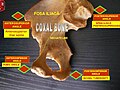
 Coxal bone (ilium, ischium and pubis).jpg 960 × 720; 87 KB
-
 Coxal bone - angles.jpg 960 × 720; 109 KB
-
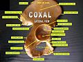
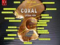
 Coxal bone - lateral view.jpg 960 × 720; 118 KB
-
 Coxal bone - medial view.jpg 960 × 720; 110 KB
-
 Coxal bone - muscular insertions.jpg 960 × 720; 94 KB
-
 Coxal bone.jpg 960 × 720; 76 KB
-
 HK TST Science Museum Bones exhibit 23 人類 skeletons.JPG 2,448 × 3,264; 1.61 MB
-
 Human hip bone texture.jpg 3,872 × 2,592; 5.09 MB
-
 Pelvis - os coxae (dex, sin).jpg 4,608 × 3,456; 5.03 MB
-
 Pelvis - os coxae (lateral view) 2.jpg 4,608 × 3,456; 4.4 MB
-
 Pelvis - os coxae (lateral view).jpg 3,456 × 4,608; 4.71 MB
-
 Pelvis - os coxae - dex, sin (lateral).jpg 4,608 × 3,456; 4.92 MB
-
 Pelvis - os illium - detail of bone tissue.jpg 4,288 × 2,704; 4.01 MB
-
 Slide12DEN.JPG 960 × 720; 100 KB
-
 Slide14DEN.JPG 960 × 720; 106 KB
-
 Slide2AA.JPG 960 × 720; 102 KB
-
 Slide3A.JPG 960 × 720; 109 KB
-
 Slide6A.JPG 960 × 720; 104 KB
-
 Small-Bodied Humans from Palau, Micronesia - Bones.png 303 × 270; 53 KB
.jpg)


.jpg)
_2.jpg)
.jpg)
.jpg)


