File:Cell-Death-in-the-Epithelia-of-the-Intestine-and-Hepatopancreas-in-Neocaridina-heteropoda-pone.0147582.s005.ogv
Size of this JPG preview of this OGG file: 800 × 450 pixels. Other resolutions: 320 × 180 pixels | 640 × 360 pixels | 1,296 × 729 pixels.
Original file (Ogg Theora video file, length 30 s, 1,296 × 729 pixels, 9.84 Mbps, file size: 35.03 MB)
Captions
Captions
Add a one-line explanation of what this file represents
Summary
edit| Description |
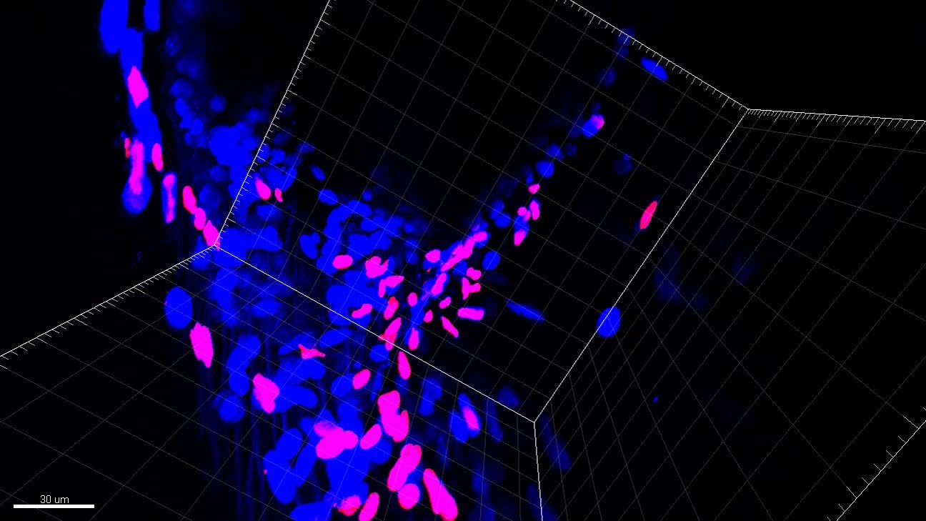
English: 3D representation of apoptotic cells in the intestine of N . heteropoda . Nuclei of apoptotic cells (red), nuclei (blue). TUNEL assay and Hoechst 33342 staining. Confocal microscope. |
||
| Date | |||
| Source | S3 Video from Sonakowska L, Włodarczyk A, Wilczek G, Wilczek P, Student S, Rost-Roszkowska M (2016). "Cell Death in the Epithelia of the Intestine and Hepatopancreas in Neocaridina heteropoda (Crustacea, Malacostraca)". PLOS ONE. DOI:10.1371/journal.pone.0147582. PMID 26844766. PMC: 4741826. | ||
| Author | Sonakowska L, Włodarczyk A, Wilczek G, Wilczek P, Student S, Rost-Roszkowska M | ||
| Permission (Reusing this file) |
This file is licensed under the Creative Commons Attribution 4.0 International license.
|
||
| Provenance |
|
File history
Click on a date/time to view the file as it appeared at that time.
| Date/Time | Thumbnail | Dimensions | User | Comment | |
|---|---|---|---|---|---|
| current | 07:43, 29 February 2016 | 30 s, 1,296 × 729 (35.03 MB) | Open Access Media Importer Bot (talk | contribs) | Automatically uploaded media file from Open Access source. Please report problems or suggestions here. |
You cannot overwrite this file.
File usage on Commons
The following page uses this file: