File:Charinus africanus (10.5852-ejt.2021.772.1505) Figure 84.png
Size of this preview: 800 × 597 pixels. Other resolutions: 320 × 239 pixels | 640 × 478 pixels | 1,024 × 765 pixels | 1,280 × 956 pixels | 1,895 × 1,415 pixels.
Original file (1,895 × 1,415 pixels, file size: 1.39 MB, MIME type: image/png)
Captions
Captions
Add a one-line explanation of what this file represents
Summary
edit_Figure_84.png&action=edit§ion=1)
| Description |
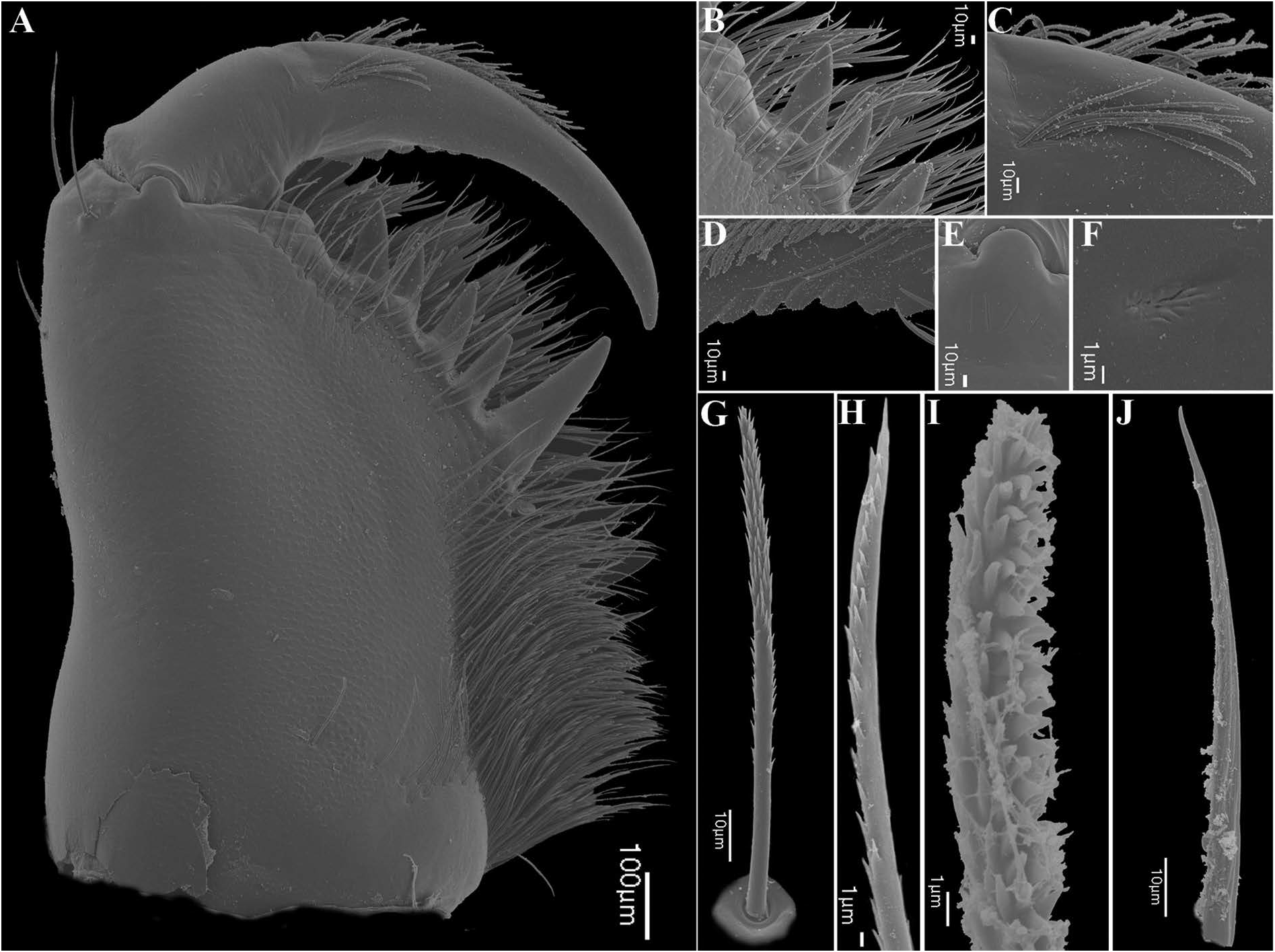
English: Fig. 84. Charinus africanus Hansen, 1921 (AMCC [LP 6943]), chelicera, ♀. A. Prolateral view. B. Retrolateral projection, opposite to bifid tooth. C. Setal row on cheliceral claw. D. Cheliceral claw teeth. E. Position of gland on joint between basal segment and cheliceral claw. F. Gland. G. Seta on cheliceral basal segment. H. Apex of basal segment seta. I. Seta on cheliceral claw. J. Setae on dorsal surface of cheliceral basal segment. |
| Date | |
| Source | https://doi.org/10.5852/ejt.2021.772.1505 |
| Author | Miranda, G. S. de, Giupponi, A. P., Prendini, L., & Scharff, N. (2021). Systematic revision of the pantropical whip spider family Charinidae Quintero, 1986 (Arachnida, Amblypygi). European Journal of Taxonomy, 772(1), 1-409. |
| Permission (Reusing this file) |
This file is licensed under the Creative Commons Attribution 4.0 International license.
|
File history
Click on a date/time to view the file as it appeared at that time.
| Date/Time | Thumbnail | Dimensions | User | Comment | |
|---|---|---|---|---|---|
| current | 11:57, 14 March 2022 | | 1,895 × 1,415 (1.39 MB) | Christian Ferrer (talk | contribs) | {{Information | description = {{en|1=Fig. 84. ''Charinus africanus'' Hansen, 1921 (AMCC [LP 6943]), chelicera, ♀. A. Prolateral view. B. Retrolateral projection, opposite to bifid tooth. C. Setal row on cheliceral claw. D. Cheliceral claw teeth. E. Position of gland on joint between basal segment and cheliceral claw. F. Gland. G. Seta on cheliceral basal segment. H. Apex of basal segment seta. I. Seta on cheliceral claw. J. Setae on dorsal surface of cheliceral basal segment.}} | date = 202... |
You cannot overwrite this file.
File usage on Commons
There are no pages that use this file.
_Figure_84.png&oldid=854432221)