File:Coarctation and PDA.png
Coarctation_and_PDA.png (555 × 568 pixels, file size: 43 KB, MIME type: image/png)
Captions
Captions
Summary
edit
| Description |
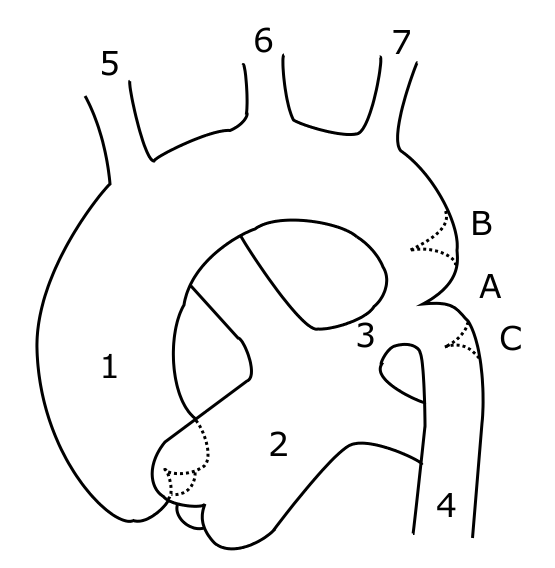
Schematic drawing of alternative locations of a coarctation of the aorta. I, Kjetil Lenes, have made the drawing myself, after information from Valdes-Cruz LM, Cayre RO: Echocardiographic diagnosis of congenital heart disease. Philadelhia, 1998.. Legend: A: ductal coarctation, B: preductal coarctation, C: postductal coarctation. 1: Aorta ascendens, 2: Arteria pulmonalis, 3: Ductus arteriosus, 4: Aorta descendens, 5: Trunchus brachiocephalicus, 6: Arteria carotis communis sinister, 7: Arteria subclavia sinister The picture is somewhat misleading, with left pulmonary artery crossing behind aorta. This will be changed in a future drawing. |
| Date | 23 May 2006 (original upload date) |
| Source | No machine-readable source provided. Own work assumed (based on copyright claims). |
| Author | No machine-readable author provided. Ekko assumed (based on copyright claims). |
Licensing
edit
| I, the copyright holder of this work, release this work into the public domain. This applies worldwide. In some countries this may not be legally possible; if so: I grant anyone the right to use this work for any purpose, without any conditions, unless such conditions are required by law. |
File history
Click on a date/time to view the file as it appeared at that time.
| Date/Time | Thumbnail | Dimensions | User | Comment | |
|---|---|---|---|---|---|
| current | 08:14, 23 May 2006 | | 555 × 568 (43 KB) | Ekko (talk | contribs) | Schematic drawing of alternative locations of a coarctation of the aorta. I, Kjetil Lenes, have made the drawing myself, after information from Valdes-Cruz LM, Cayre RO: ''Echocardiographic diagnosis of congenital heart disease.'' Philadelhia, 1998.. Lege |
You cannot overwrite this file.
File usage on Commons
There are no pages that use this file.
File usage on other wikis
The following other wikis use this file:
- Usage on ar.wikipedia.org
- Usage on es.wikipedia.org
- Usage on fr.wikipedia.org
- Usage on it.wikipedia.org
- Usage on nn.wikipedia.org
- Usage on no.wikipedia.org
- Usage on pl.wikipedia.org
- Usage on pt.wikipedia.org
- Usage on sr.wikipedia.org
- Usage on uk.wikipedia.org
- Usage on www.wikidata.org