File:Craniofacial-divergence-by-distinct-prenatal-growth-patterns-in-Fgfr2-mutant-mice-1471-213X-14-8-S4.ogv
Size of this JPG preview of this OGG file: 800 × 453 pixels. Other resolutions: 320 × 181 pixels | 640 × 362 pixels | 848 × 480 pixels.
Original file (Ogg Theora video file, length 0.0 s, 848 × 480 pixels, 0 bps, file size: 15.98 MB)
Captions
Captions
Add a one-line explanation of what this file represents
Summary
edit| Description |
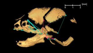
English: Apert253_E17.5_EDMA. Video 2: 3D reconstruction of isosurface of E17.5 mouse skull showing linear differences in cranial morphology as measured between 3D coordinates of anatomical landmarks between Fgfr2
+/P253R mice and unaffected littermates following Figure 4E and F. Blue lines are significantly larger in mutant mice relative to unaffected littermates. Fuchsia lines are significantly smaller in mutant mice. Bone segmented from HRμCT images is shown as partially transparent to better visualize the growth differences. |
||
| Date | |||
| Source | Video file from Motch Perrine S, Cole T, Martinez-Abadias N, Aldridge K, Jabs E, Richtsmeier J (2014). "Craniofacial divergence by distinct prenatal growth patterns in Fgfr2 mutant mice". BMC Developmental Biology. DOI:10.1186/1471-213X-14-8. PMID 24580805. PMC: 4101838. | ||
| Author | Motch Perrine S, Cole T, Martinez-Abadias N, Aldridge K, Jabs E, Richtsmeier J | ||
| Permission (Reusing this file) |
This file is licensed under the Creative Commons Attribution 2.0 Generic license.
|
||
| Provenance |
|
File history
Click on a date/time to view the file as it appeared at that time.
| Date/Time | Thumbnail | Dimensions | User | Comment | |
|---|---|---|---|---|---|
| current | 03:35, 21 July 2014 | 0.0 s, 848 × 480 (15.98 MB) | Open Access Media Importer Bot (talk | contribs) | Automatically uploaded media file from Open Access source. Please report problems or suggestions here. |
You cannot overwrite this file.
File usage on Commons
There are no pages that use this file.