File:Experimental-Cerebral-Malaria-Pathogenesis—Hemodynamics-at-the-Blood-Brain-Barrier-ppat.1004528.s025.ogv
Size of this JPG preview of this OGG file: 667 × 600 pixels. Other resolutions: 267 × 240 pixels | 534 × 480 pixels | 806 × 725 pixels.
Original file (Ogg Theora video file, length 10 s, 806 × 725 pixels, 2.81 Mbps, file size: 3.35 MB)
Captions
Captions
Add a one-line explanation of what this file represents
Summary
edit| Description |
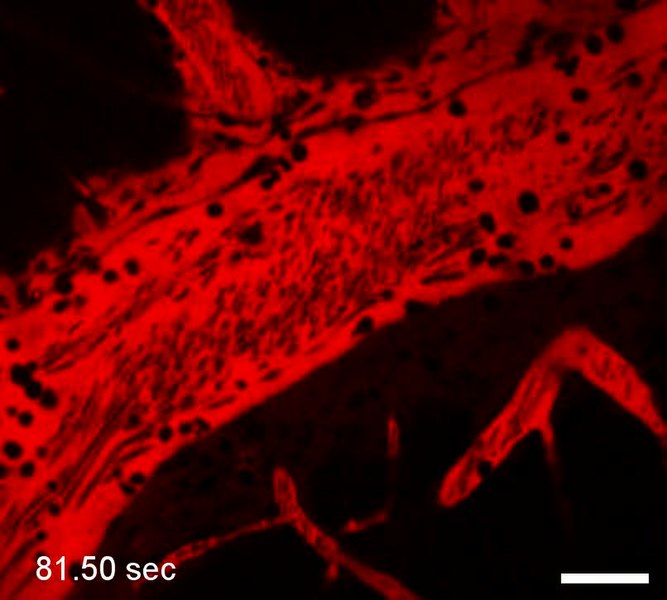
English: ECM is associated with a significant reduction in the venous blood flow. Intravital microscopy of a PbA-infected CBA/CaJ mouse injected with Evans blue (red) upon development of ECM symptoms. Arrested leukocytes (dark circles) prevent access to the endothelium so that the marginal zone of the postcapillary venule lumen lacks blood cells traveling in the bloodstream (dark streaks). Evans blue has leaked from the cortical postcapillary venules in the surrounding tissue. Scale bar = 20 µm. |
||
| Date | |||
| Source | Video S2 from Nacer A, Movila A, Sohet F, Girgis N, Gundra U, Loke P, Daneman R, Frevert U (2014). "Experimental Cerebral Malaria Pathogenesis—Hemodynamics at the Blood Brain Barrier". PLOS Pathogens. DOI:10.1371/journal.ppat.1004528. PMID 25474413. PMC: 4256476. | ||
| Author | Nacer A, Movila A, Sohet F, Girgis N, Gundra U, Loke P, Daneman R, Frevert U | ||
| Permission (Reusing this file) |
This file is licensed under the Creative Commons Attribution 4.0 International license.
|
||
| Provenance |
|
File history
Click on a date/time to view the file as it appeared at that time.
| Date/Time | Thumbnail | Dimensions | User | Comment | |
|---|---|---|---|---|---|
| current | 22:50, 13 December 2014 | 10 s, 806 × 725 (3.35 MB) | Open Access Media Importer Bot (talk | contribs) | Automatically uploaded media file from Open Access source. Please report problems or suggestions here. |
You cannot overwrite this file.
File usage on Commons
There are no pages that use this file.