File:Experimental-Cerebral-Malaria-Pathogenesis—Hemodynamics-at-the-Blood-Brain-Barrier-ppat.1004528.s035.ogv
Size of this JPG preview of this OGG file: 650 × 600 pixels. Other resolutions: 260 × 240 pixels | 520 × 480 pixels | 715 × 660 pixels.
Original file (Ogg Theora video file, length 5.0 s, 715 × 660 pixels, 3.36 Mbps, file size: 2 MB)
Captions
Captions
Add a one-line explanation of what this file represents
Summary
edit| Description |
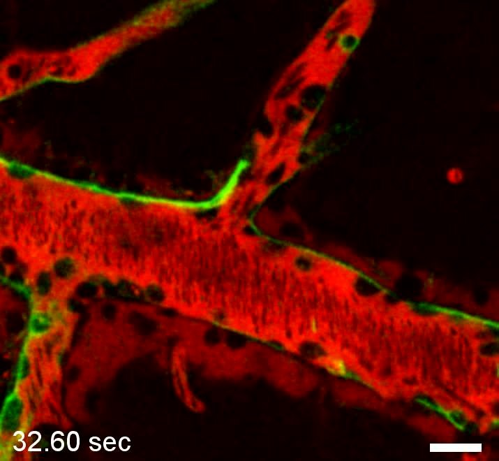
English: Monocyte behavior during ECM. Intravital microscopy of a PbA infected CBA/CaJ mouse with ECM showing monocytes (green surface label) arrested on the endothelium of a cortical postcapillary venules. Monocytes were labeled by intravenous inoculation of PE-conjugated anti-CD14. Note that the fluorescent marker also labels the endothelium of the postcapillary venules. The vascular lumen was visualized with Evans blue (red). The numerous large leukocytes (dark circles) represent most likely monocytes and macrophages. Scale bar = 20 µm. |
||
| Date | |||
| Source | Video S12 from Nacer A, Movila A, Sohet F, Girgis N, Gundra U, Loke P, Daneman R, Frevert U (2014). "Experimental Cerebral Malaria Pathogenesis—Hemodynamics at the Blood Brain Barrier". PLOS Pathogens. DOI:10.1371/journal.ppat.1004528. PMID 25474413. PMC: 4256476. | ||
| Author | Nacer A, Movila A, Sohet F, Girgis N, Gundra U, Loke P, Daneman R, Frevert U | ||
| Permission (Reusing this file) |
This file is licensed under the Creative Commons Attribution 4.0 International license.
|
||
| Provenance |
|
File history
Click on a date/time to view the file as it appeared at that time.
| Date/Time | Thumbnail | Dimensions | User | Comment | |
|---|---|---|---|---|---|
| current | 22:52, 13 December 2014 | 5.0 s, 715 × 660 (2 MB) | Open Access Media Importer Bot (talk | contribs) | Automatically uploaded media file from Open Access source. Please report problems or suggestions here. |
You cannot overwrite this file.
File usage on Commons
There are no pages that use this file.