File:Gross-Morphological-Features-of-the-Organ-Surface-Primo-Vascular-System-Revealed-by-Hemacolor-350815.f1.ogv
Size of this JPG preview of this OGG file: 800 × 450 pixels. Other resolutions: 320 × 180 pixels | 640 × 360 pixels | 1,280 × 720 pixels.
Original file (Ogg Theora video file, length 13 s, 1,280 × 720 pixels, 479 kbps, file size: 782 KB)
Captions
Captions
Add a one-line explanation of what this file represents
Summary
edit| Description |
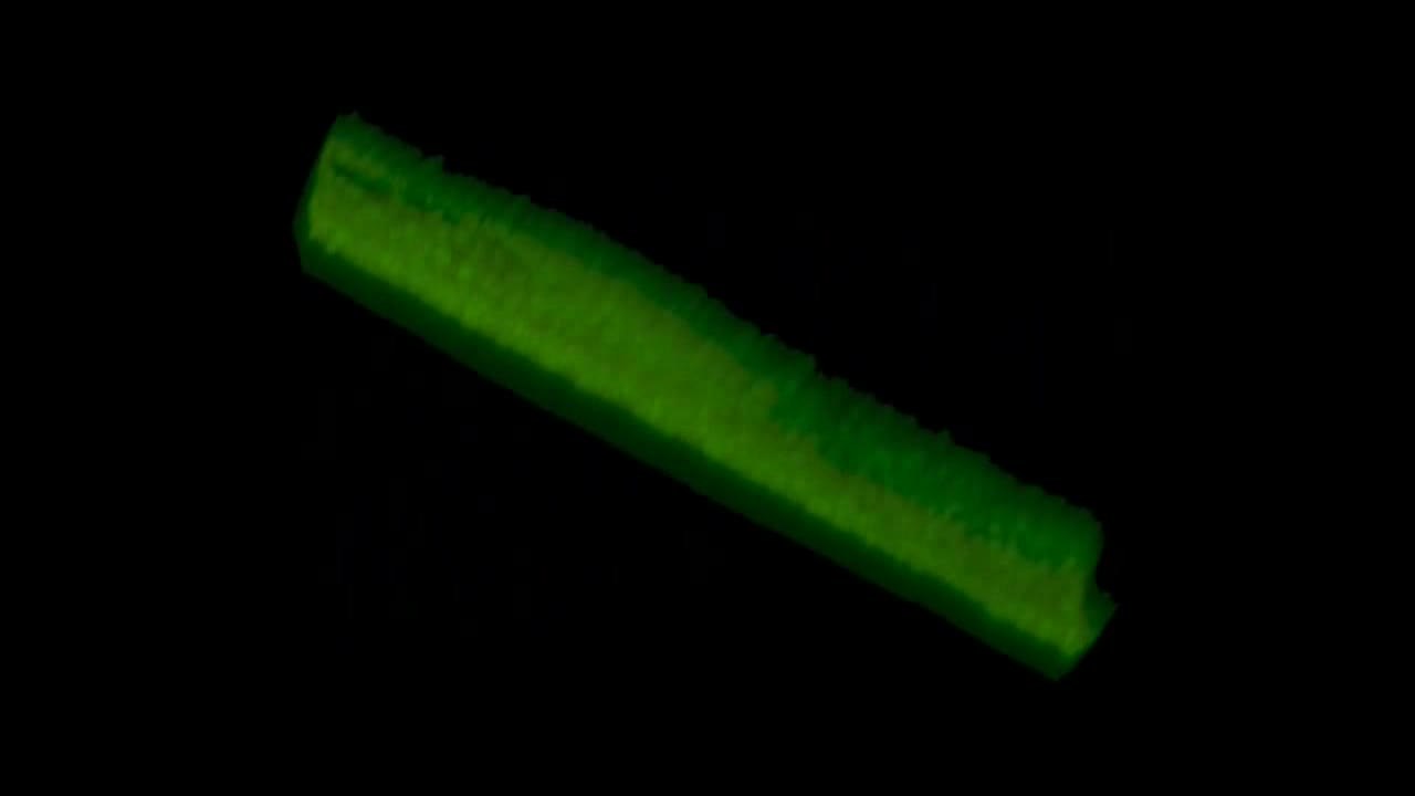
English: Movie S1: Whole primo-vessel tissue was stained with acridine orange and the 3D microscopy was performed by a confocal laser scanning microscope (LSM710, Carl Zeiss, Germany) to show an inner space part within the primo-vessel. Most cells of the primo-vessel were stained green color (denoting DNA), and the inner space with low cellularity appeared dark along the longitudinal axis of the primo-vessel.
Movie S2: Spontaneous movement of granules within a putative mast cell in the primo-node slice was recorded under a light microscope with differential interference contrast (BX50WI, Olympus, Tokyo, Japan) using a USB digital CCD camera series 150PIII. Movie S3: Spontaneous movement of a granule in isolation was recorded under a light microscope with differential interference contrast (BX50WI, Olympus, Tokyo, Japan) using a USB digital CCD camera series 150PIII. |
||
| Date | |||
| Source | Lim C, Yoo J, Kim Y, Lee S, Ryu P (2013). "Gross Morphological Features of the Organ Surface Primo-Vascular System Revealed by Hemacolor Staining". Evidence-based Complementary and Alternative Medicine. DOI:10.1155/2013/350815. PMID 23986781. PMC: 3748414. | ||
| Author | Lim C, Yoo J, Kim Y, Lee S, Ryu P | ||
| Permission (Reusing this file) |
This file is licensed under the Creative Commons Attribution 3.0 Unported license.
|
||
| Provenance |
|
File history
Click on a date/time to view the file as it appeared at that time.
| Date/Time | Thumbnail | Dimensions | User | Comment | |
|---|---|---|---|---|---|
| current | 22:10, 4 September 2013 | 13 s, 1,280 × 720 (782 KB) | Open Access Media Importer Bot (talk | contribs) | Automatically uploaded media file from Open Access source. Please report problems or suggestions here. |
You cannot overwrite this file.
File usage on Commons
There are no pages that use this file.