File:Imaging-of-human-differentiated-3D-neural-aggregates-using-light-sheet-fluorescence-microscopy-Movie1.ogv
Size of this JPG preview of this OGG file: 800 × 400 pixels. Other resolutions: 320 × 160 pixels | 830 × 415 pixels.
Original file (Ogg Theora video file, length 12 s, 830 × 415 pixels, 523 kbps, file size: 766 KB)
Captions
Captions
Add a one-line explanation of what this file represents
Summary
edit| Description |
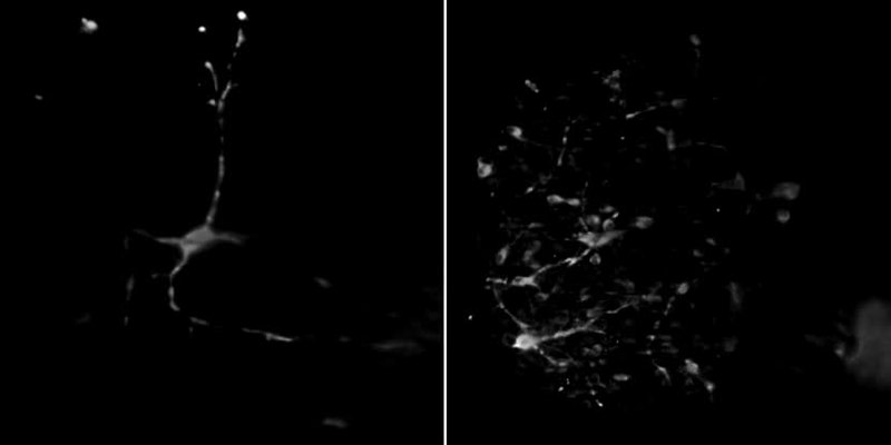
English: 3D reconstructions of two dopaminergic neuronal networks stained with the dopaminergic marker tyrosine hydroxylase (TH) showing their maturation and functionality. |
||
| Date | |||
| Source | Movie S1 from Gualda E, Simao D, Pinto C, Alves P, Brito C (2014). "Imaging of human differentiated 3D neural aggregates using light sheet fluorescence microscopy". Frontiers in Cellular Neuroscience. DOI:10.3389/fncel.2014.00221. PMID 25161607. PMC: 4123789. | ||
| Author | Gualda E, Simao D, Pinto C, Alves P, Brito C | ||
| Permission (Reusing this file) |
This file is licensed under the Creative Commons Attribution 3.0 Unported license.
|
||
| Provenance |
|
File history
Click on a date/time to view the file as it appeared at that time.
| Date/Time | Thumbnail | Dimensions | User | Comment | |
|---|---|---|---|---|---|
| current | 23:35, 28 August 2014 | 12 s, 830 × 415 (766 KB) | Open Access Media Importer Bot (talk | contribs) | Automatically uploaded media file from Open Access source. Please report problems or suggestions here. |
You cannot overwrite this file.
File usage on Commons
There are no pages that use this file.