File:Left-and-Right-Lung-Asynchrony-as-a-Physiological-Indicator-for-Unilateral-Bronchial-Obstruction-in-pone.0105327.s002.ogv
Size of this JPG preview of this OGG file: 800 × 450 pixels. Other resolutions: 320 × 180 pixels | 640 × 360 pixels | 960 × 540 pixels.
Original file (Ogg Theora video file, length 20 s, 960 × 540 pixels, 217 kbps, file size: 526 KB)
Captions
Captions
Add a one-line explanation of what this file represents
Summary
edit| Description |
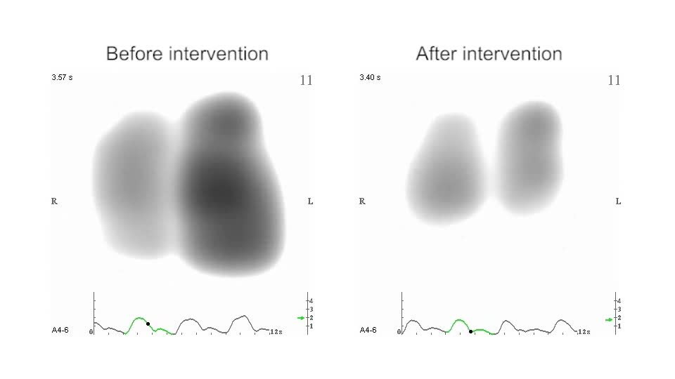
English: Lung sound distribution (2). Left panel: VRI before intervention. Right panel: VRI after intervention. The development of lung sound intensity in the left lung lagged behind the right. After intervention, these asynchronies were mostly resolved. The biphasic curve at the bottom stands for inspiratory and expiratory lung sound intensity. |
||
| Date | |||
| Source | Video S2 from Mineshita M, Kida H, Nishine H, Handa H, Inoue T, Miyazawa T (2014). "Left and Right Lung Asynchrony as a Physiological Indicator for Unilateral Bronchial Obstruction in Interventional Bronchoscopy". PLOS ONE. DOI:10.1371/journal.pone.0105327. PMID 25133760. PMC: 4136828. | ||
| Author | Mineshita M, Kida H, Nishine H, Handa H, Inoue T, Miyazawa T | ||
| Permission (Reusing this file) |
This file is licensed under the Creative Commons Attribution 4.0 International license.
|
||
| Provenance |
|
File history
Click on a date/time to view the file as it appeared at that time.
| Date/Time | Thumbnail | Dimensions | User | Comment | |
|---|---|---|---|---|---|
| current | 07:50, 25 August 2014 | 20 s, 960 × 540 (526 KB) | Open Access Media Importer Bot (talk | contribs) | Automatically uploaded media file from Open Access source. Please report problems or suggestions here. |
You cannot overwrite this file.
File usage on Commons
There are no pages that use this file.