File:Mapping-the-dynamics-and-nanoscale-organization-of-synaptic-adhesion-proteins-using-monomeric-ncomms10773-s5.ogv
Size of this JPG preview of this OGG file: 774 × 600 pixels. Other resolutions: 310 × 240 pixels | 620 × 480 pixels | 992 × 768 pixels | 1,158 × 897 pixels.
Original file (Ogg Theora video file, length 21 s, 1,158 × 897 pixels, 10.45 Mbps, file size: 25.79 MB)
Captions
Captions
Add a one-line explanation of what this file represents
Summary edit
| Description |
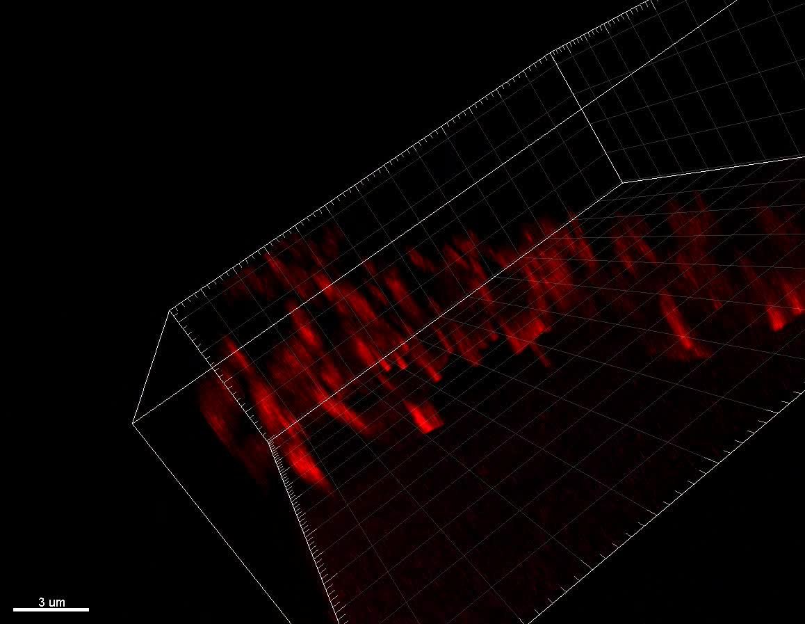
English: Supplementary Movie 4 STED imaging of neuroligin-1 accumulation in dendritic spines. High magnification deconvolved STED (red) and confocal (GFP) 3D animations from a dendritic portion of a single cell electroporated neuron expressing GFP, AP-Nlg1, and BirAER, in a mouse organotypic brain slice. Note the strong labeling of Nlg1 in dendritic spines. |
||
| Date | |||
| Source | Video file from Chamma I, Letellier M, Butler C, Tessier B, Lim K, Gauthereau I, Choquet D, Sibarita J, Park S, Sainlos M, Thoumine O (2016). "Mapping the dynamics and nanoscale organization of synaptic adhesion proteins using monomeric streptavidin". Nature Communications. DOI:10.1038/ncomms10773. PMID 26979420. PMC: 4799371. | ||
| Author | Chamma I, Letellier M, Butler C, Tessier B, Lim K, Gauthereau I, Choquet D, Sibarita J, Park S, Sainlos M, Thoumine O | ||
| Permission (Reusing this file) |
This file is licensed under the Creative Commons Attribution 4.0 International license.
|
||
| Provenance |
|
File history
Click on a date/time to view the file as it appeared at that time.
| Date/Time | Thumbnail | Dimensions | User | Comment | |
|---|---|---|---|---|---|
| current | 17:11, 28 October 2016 | 21 s, 1,158 × 897 (25.79 MB) | Open Access Media Importer Bot (talk | contribs) | Automatically uploaded media file from Open Access source. Please report problems or suggestions here. |
You cannot overwrite this file.
File usage on Commons
There are no pages that use this file.