File:Meningeal-cells-and-glia-establish-a-permissive-environment-for-axon-regeneration-after-spinal-cord-1749-8104-6-1-S10.ogv
Size of this JPG preview of this OGG file: 355 × 600 pixels. Other resolutions: 142 × 240 pixels | 606 × 1,024 pixels.
Original file (Ogg Theora video file, length 12 s, 606 × 1,024 pixels, 2.28 Mbps, file size: 3.23 MB)
Captions
Captions
Add a one-line explanation of what this file represents
Summary
edit| Description |
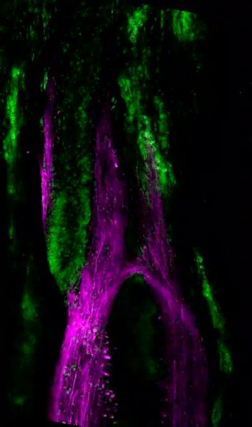
English: Movie 6: movie through confocal z-stack of animal shown in Additional file 8E, a non-recovered animal. This is a 9-week regenerate in the growth beyond stage that had not recovered function. It begins on the ventral side of the cord and moves in 2-μm increments through to the dorsal side. Rostral is up. Descending axons are labeled with the axon tracer in magenta and nuclei are in green. Note there are two ependymal tubes on the rostral side. |
||
| Date | |||
| Source | Zukor K, Kent D, Odelberg S (2011). "Meningeal cells and glia establish a permissive environment for axon regeneration after spinal cord injury in newts". Neural Development. DOI:10.1186/1749-8104-6-1. PMID 21205291. PMC: 3025934. | ||
| Author | Zukor K, Kent D, Odelberg S | ||
| Permission (Reusing this file) |
This file is licensed under the Creative Commons Attribution 2.0 Generic license.
|
||
| Provenance |
|
File history
Click on a date/time to view the file as it appeared at that time.
| Date/Time | Thumbnail | Dimensions | User | Comment | |
|---|---|---|---|---|---|
| current | 03:29, 20 November 2012 | 12 s, 606 × 1,024 (3.23 MB) | Open Access Media Importer Bot (talk | contribs) | Automatically uploaded media file from Open Access source. Please report problems or suggestions here. |
You cannot overwrite this file.
File usage on Commons
There are no pages that use this file.