File:Neanthes goodayi (10.5852-ejt.2021.760.1447) Figure 2.png

Size of this preview: 462 × 600 pixels. Other resolutions: 185 × 240 pixels | 370 × 480 pixels | 592 × 768 pixels | 789 × 1,024 pixels | 1,895 × 2,459 pixels.
Original file (1,895 × 2,459 pixels, file size: 6.65 MB, MIME type: image/png)
Captions
Captions
Add a one-line explanation of what this file represents
Summary
edit| Description |
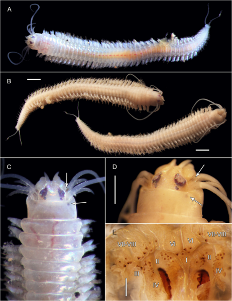
English: Fig. 2. Neanthes goodayi sp. nov., holotype (NHM_739). A. Live image, entire specimen. B. Preserved entire specimen, dorsal view (left), ventral view (right). C. Live image, anterior view, arrows mark pigmentation. D. Preserved specimen, anterior view, arrows mark pigmentation. E. Dissected pharynx, with pharyngeal areas I, II, III, IV, VI, VII–VIII highlighted. Scale bars: B = 1 mm; D = 500 µm; E = 250 µm. |
| Date | |
| Source | https://doi.org/10.5852/ejt.2021.760.1447 |
| Author | Drennan, R., Wiklund, H., Rabone, M., Georgieva, M. N., Dahlgren, T. G., & Glover, A. G. (2021). Neanthes goodayi sp. nov. (Annelida, Nereididae), a remarkable new annelid species living inside deep-sea polymetallic nodules. European Journal of Taxonomy, 760(1), 160-185. |
| Permission (Reusing this file) |
This file is licensed under the Creative Commons Attribution 4.0 International license.
|
| Other versions |
_Figure_2_(cropped).png)
_Figure_2.png&action=edit§ion=1)
File history
Click on a date/time to view the file as it appeared at that time.
| Date/Time | Thumbnail | Dimensions | User | Comment | |
|---|---|---|---|---|---|
| current | 07:47, 26 June 2022 | | 1,895 × 2,459 (6.65 MB) | Christian Ferrer (talk | contribs) | {{Information | description = {{en|1=Fig. 2. ''Neanthes goodayi'' sp. nov., holotype (NHM_739). A. Live image, entire specimen. B. Preserved entire specimen, dorsal view (left), ventral view (right). C. Live image, anterior view, arrows mark pigmentation. D. Preserved specimen, anterior view, arrows mark pigmentation. E. Dissected pharynx, with pharyngeal areas I, II, III, IV, VI, VII–VIII highlighted. Scale bars: B = 1 mm; D = 500 µm; E = 250 µm.}} | date = 2021-07-27 | source = https://d... |
You cannot overwrite this file.
File usage on Commons
The following page uses this file:
_Figure_2.png&oldid=668577763)