File:Notaulax tenuitorques (10.7717-peerj.9692) Fig-11-full.png

Size of this preview: 737 × 600 pixels. Other resolutions: 295 × 240 pixels | 590 × 480 pixels | 944 × 768 pixels | 1,258 × 1,024 pixels | 1,619 × 1,318 pixels.
Original file (1,619 × 1,318 pixels, file size: 2.08 MB, MIME type: image/png)
Captions
Captions
Add a one-line explanation of what this file represents
Summary
edit_Fig-11-full.png&action=edit§ion=1)
| Description |
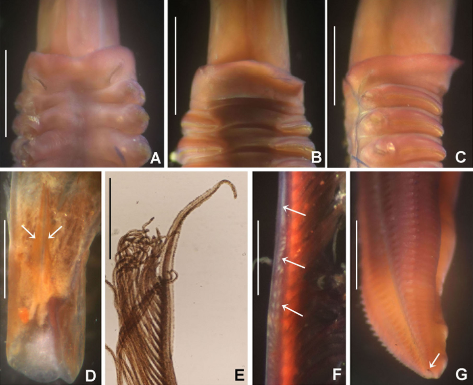
English: Figure 11: Notaulax tenuitorques.
|
| Date | |
| Source | https://doi.org/10.7717/peerj.9692 |
| Author | Tovar-Hernández MA, ten Hove HA, Vinn O, Zatoń M, de León-González JA, García-Garza ME. 2020. Fan worms (Annelida: Sabellidae) from Indonesia collected by the Snellius II Expedition (1984) with descriptions of three new species and tube microstructure. PeerJ 8: e9692 |
| Permission (Reusing this file) |
This file is licensed under the Creative Commons Attribution 4.0 International license.
|
File history
Click on a date/time to view the file as it appeared at that time.
| Date/Time | Thumbnail | Dimensions | User | Comment | |
|---|---|---|---|---|---|
| current | 18:20, 2 January 2022 | | 1,619 × 1,318 (2.08 MB) | Christian Ferrer (talk | contribs) | {{Information |description={{en|1= Figure 11: ''Notaulax tenuitorques''. :(A) Collar and first thoracic chaetigers, dorsal view, (B) same, ventral view, (C) same, lateral view, (D) dorsal lips indicated by arrows, (E) radiolar tip, (F) radiolar ocelli indicated by arrows, (G) posterior abdomen and pygidial eye indicated by arrow. Scale bars: (A–C) 1.5 mm, (D–E, G) 1 mm, (F) 0.3 mm. Stain: (A–C, F–G) shirla. Single specimens, RMNH.VER. 19941.}} |date= 2020-08-18 |source=https://doi.org/10.7717... |
You cannot overwrite this file.
File usage on Commons
The following page uses this file:
File usage on other wikis
The following other wikis use this file:
- Usage on species.wikimedia.org
- Usage on www.wikidata.org
_Fig-11-full.png&oldid=618634746)