File:Ophiogaleus (10.5852-ejt.2018.411) Figure 5.png

Size of this preview: 439 × 599 pixels. Other resolutions: 176 × 240 pixels | 351 × 480 pixels | 562 × 768 pixels | 750 × 1,024 pixels | 1,683 × 2,298 pixels.
Original file (1,683 × 2,298 pixels, file size: 5.92 MB, MIME type: image/png)
Captions
Captions
Add a one-line explanation of what this file represents
Summary
edit_Figure_5.png&action=edit§ion=1)
| Description |
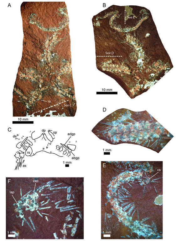
English: Fig. 5. Ophiogaleus sp. A–E. PMO 218.060, specimen preserving the ventral side of the disc area and the base of all five arms. The specimen has been split through a horizontal plane and is visible as part (A) and counterpart (B). Photographs in A and B by H.A. Nakrem. C. Drawing detail of the oral and interradial area. D. Detail view of proximal arm segments with articulated spines. E. Detail view of distal arm segment. F. PMO 218.053a, partly disarticulated arm portion composed of three median segments showing details of the ventral side of the arms. Abbreviations: abgp = abradial genital plate; adgp = adradial genital plate; as = arm spine; cs = arm in cross section; dp = dental plate; ds = disc spine; lap = lateral arm plate; op = oral plate; rs = radial shield; vap = ventral arm plate. |
| Date | |
| Source | Rousseau, J., Gale, A. S., & Thuy, B. (2018). New articulated asteroids (Echinodermata, Asteroidea) and ophiuroids (Echinodermata, Ophiuroidea) from the Late Jurassic (Volgian / Tithonian) of central Spitsbergen. European Journal of Taxonomy, (411). https://doi.org/10.5852/ejt.2018.411 |
| Author | Rousseau, Gale & Thuy (2018) |
| Permission (Reusing this file) |
This file is licensed under the Creative Commons Attribution 3.0 Unported license.
|
File history
Click on a date/time to view the file as it appeared at that time.
| Date/Time | Thumbnail | Dimensions | User | Comment | |
|---|---|---|---|---|---|
| current | 05:32, 5 August 2023 | | 1,683 × 2,298 (5.92 MB) | Christian Ferrer (talk | contribs) | {{Information | description = {{en|1=Fig. 5. ''Ophiogaleus'' sp. A–E. PMO 218.060, specimen preserving the ventral side of the disc area and the base of all five arms. The specimen has been split through a horizontal plane and is visible as part (A) and counterpart (B). Photographs in A and B by H.A. Nakrem. C. Drawing detail of the oral and interradial area. D. Detail view of proximal arm segments with articulated spines. E. Detail view of distal arm segment. F. PMO 218.053a, partly disarti... |
You cannot overwrite this file.
File usage on Commons
The following 2 pages use this file:
_Figure_5.png&oldid=790960068)