File:Plate 1 (Taylor, 1945).jpg
Size of this preview: 391 × 600 pixels. Other resolutions: 156 × 240 pixels | 313 × 480 pixels | 644 × 988 pixels.
Original file (644 × 988 pixels, file size: 57 KB, MIME type: image/jpeg)
Captions
Captions
Add a one-line explanation of what this file represents
Summary
edit.jpg&action=edit§ion=1)
| Description |
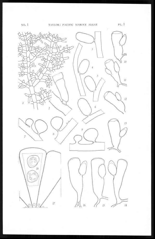
English: Plate 1. Figs. 1, 2. Boodlea composita, f. Fig. 1, a small portion of a thallus to show its characteristics in the plane phase, x 15. Fig. 2, tip of a branchlet showing a hapteral cell attaching to a larger segment, x 73. Ecuador. Figs. 3-6. Derbesia longifructa. Four oval-oblong sporangia attached to the supporting filaments, showing the basal septum or, in fig. 3, a definite stalk cell. Ecuador, x 115. Figs. 7-9. Derbesia Hollenbergii. Portions of three filaments to show young subturbinate sporangia, and an old sporangium with an evident stalk cell. I. Santa Maria, x 70. Figs. 10-13. Codium isabelae. Four peripheral utricles, each showing a much-thickened end wall and one or two gametangia. I. Isabela. x 70. Figs. 14-16. Codium santamariae. Three peripheral utricles with slightly thickened end walls and solitaria gametangia. I. Santa Maria, x 70. [= syn. of Codium isabelae, AlgaeBase] Figs. 17-18. Acetabularia parvula v. americana. Fig. 17, one segment of a disk and portions of others, to show the unarmed end wall, x 90. Fig. 18, insert, two coronal processes to show the hair scars, x 140. Is. Revilla Gigedo. [= syn. of Parvocaulis parvulus, AlgaeBase] |
| Date | |
| Source | http://digitallibrary.usc.edu/cdm/compoundobject/collection/p15799coll82/id/22673 |
| Author | Taylor, William Randolph 1945. Pacific marine algae of the Allan Hancock Expeditions to the Galapagos Islands. Allan Hancock Pacific Expeditions; v. 12. University of Southern California. |
| Permission (Reusing this file) |
This file is licensed under the Creative Commons Attribution 4.0 International license.
|
This image was intentionally uploaded with a border. Please do not remove the border. Upload a new version under a different filename without a border if you want a version without a border.
|
File history
Click on a date/time to view the file as it appeared at that time.
| Date/Time | Thumbnail | Dimensions | User | Comment | |
|---|---|---|---|---|---|
| current | 17:58, 27 November 2019 | | 644 × 988 (57 KB) | Christian Ferrer (talk | contribs) | {{Information |description={{en|1=Plate 1. <br>Figs. 1, 2. ''Boodlea composita, f''. Fig. 1, a small portion of a thallus to show its characteristics in the plane phase, x 15. Fig. 2, tip of a branchlet showing a hapteral cell attaching to a larger segment, x 73. Ecuador.<br>Figs. 3-6. ''Derbesia longifructa''. Four oval-oblong sporangia attached to the supporting filaments, showing the basal septum or, in fig. 3, a definite stalk cell. Ecuador, x 115.<br>Figs. 7-9. ''Derbesia Hollenbegii''.... |
You cannot overwrite this file.
File usage on Commons
The following page uses this file:
File usage on other wikis
The following other wikis use this file:
- Usage on species.wikimedia.org
.jpg&oldid=634489527)