File:Separase-Is-Required-for-Homolog-and-Sister-Disjunction-during-Drosophila-melanogaster-Male-Meiosis-pgen.1005996.s017.ogv
Size of this JPG preview of this OGG file: 800 × 427 pixels. Other resolutions: 320 × 171 pixels | 640 × 342 pixels | 1,024 × 547 pixels | 1,648 × 880 pixels.
Original file (Ogg Theora video file, length 8.9 s, 1,648 × 880 pixels, 1.41 Mbps, file size: 1.49 MB)
Captions
Captions
Add a one-line explanation of what this file represents
Summary
edit| Description |
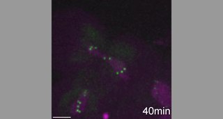
English: Normal biorientation of bivalents followed by separation failure during meiosis I after THR depletion. Time-lapse analysis of progression through the first meiotic division in spermatocytes expressing cid-EGFP and His2Av-mRFP. Moreover, spermatocyte-specific THR depletion was induced by transgenic RNAi (thr-RNAi). The movie starts at the nuclear envelope breakdown. In prometaphase I, up to 4 DNA masses, each with two green centromere dots, are recognizable. Bivalents align into a metaphase I plate indicating that spindles and kinetochores are functional. However, homologous chromosomes fail to separate during anaphase I. Precise genotype description is given in S1 Table. Image stacks with 30 focal planes spaced by 1 μm were acquired with a time interval of 1 min. Scale bar = 5 μm. |
||
| Date | |||
| Source | S6 Movie from Blattner A, Chaurasia S, McKee B, Lehner C (2016). "Separase Is Required for Homolog and Sister Disjunction during Drosophila melanogaster Male Meiosis, but Not for Biorientation of Sister Centromeres". PLOS Genetics. DOI:10.1371/journal.pgen.1005996. PMID 27120695. PMC: 4847790. | ||
| Author | Blattner A, Chaurasia S, McKee B, Lehner C | ||
| Permission (Reusing this file) |
This file is licensed under the Creative Commons Attribution 4.0 International license.
|
||
| Provenance |
|
File history
Click on a date/time to view the file as it appeared at that time.
| Date/Time | Thumbnail | Dimensions | User | Comment | |
|---|---|---|---|---|---|
| current | 01:29, 18 October 2016 | 8.9 s, 1,648 × 880 (1.49 MB) | Open Access Media Importer Bot (talk | contribs) | Automatically uploaded media file from Open Access source. Please report problems or suggestions here. |
You cannot overwrite this file.
File usage on Commons
The following page uses this file: