File:Sibogasyrinx (10.5852-ejt.2021.773.1509) Figure 8.png
Size of this preview: 490 × 600 pixels. Other resolutions: 196 × 240 pixels | 392 × 480 pixels | 627 × 768 pixels | 836 × 1,024 pixels | 1,868 × 2,287 pixels.
Original file (1,868 × 2,287 pixels, file size: 4.17 MB, MIME type: image/png)
Captions
Captions
Add a one-line explanation of what this file represents
Summary
edit_Figure_8.png&action=edit§ion=1)
| Description |
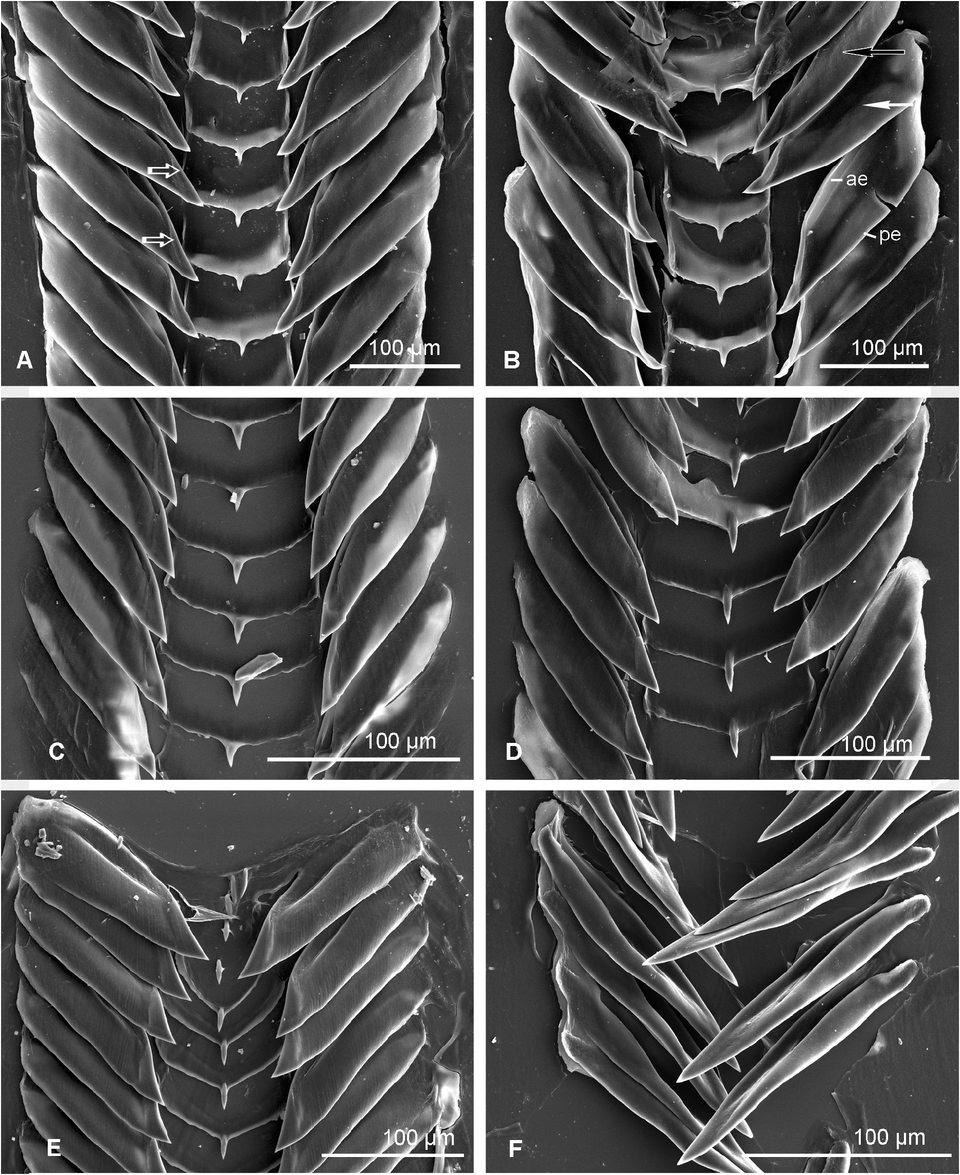
English: Fig. 8. Radulae of species of Sibogasyrinx Powell, 1969. A–B. Sibogasyrinx sangeri Kantor, Fedosov & Puillandre, 2018, MNHN-IM-2009-16995 (shell seen on Fig. 7C). A. Part of radula with fully formed marginal teeth; white hollow arrow indicates overlapping edges of the tooth at its tip. B. Part of radula showing transition between unfolded (white arrow) and completely longitudinally folded (black arrow) marginal teeth (ae = anterior tooth edge; pe = posterior tooth edge). C. Sibogasyrinx filosa Ardovini, 2021, Solomon Islands, MNHN-IM-2009-16831 (shell seen Fig. 9E–F). D. Sibogasyrinx lolae sp. nov., MNHN-IM-2009-29311. E. Sibogasyrinx maximei sp. nov., holotype, MNHN-IM-2013-45883, anterior end of radula. F. Sibogasyrinx pagodiformis sp. nov., MNHN-IM-2009-11327. |
| Date | |
| Source | https://doi.org/10.5852/ejt.2021.773.1509 |
| Author | Kantor, Y. I., & Puillandre, N. (2021). Rare, deep-water and similar: revision of Sibogasyrinx (Conoidea: Cochlespiridae). European Journal of Taxonomy, 773(1), 19-60. |
| Permission (Reusing this file) |
This file is licensed under the Creative Commons Attribution 4.0 International license.
|
File history
Click on a date/time to view the file as it appeared at that time.
| Date/Time | Thumbnail | Dimensions | User | Comment | |
|---|---|---|---|---|---|
| current | 18:06, 23 February 2022 | | 1,868 × 2,287 (4.17 MB) | Christian Ferrer (talk | contribs) | {{Information | description = {{en|1=Fig. 8. Radulae of species of ''Sibogasyrinx'' Powell, 1969. A–B. ''Sibogasyrinx sangeri'' Kantor, Fedosov & Puillandre, 2018, MNHN-IM-2009-16995 (shell seen on Fig. 7C). A. Part of radula with fully formed marginal teeth; white hollow arrow indicates overlapping edges of the tooth at its tip. B. Part of radula showing transition between unfolded (white arrow) and completely longitudinally folded (black arrow) marginal teeth (ae = anterior tooth edge; pe... |
You cannot overwrite this file.
File usage on Commons
There are no pages that use this file.
_Figure_8.png&oldid=646372165)