File:The-first-three-dimensional-visualization-of-a-thrombus-in-transit-trapped-between-the-leads-of-a-1752-1947-4-359-S3.ogv
Size of this JPG preview of this OGG file: 750 × 600 pixels. Other resolutions: 300 × 240 pixels | 600 × 480 pixels | 760 × 608 pixels.
Original file (Ogg Theora video file, length 1.8 s, 760 × 608 pixels, 2.01 Mbps, file size: 434 KB)
Captions
Captions
Add a one-line explanation of what this file represents
Summary
edit| Description |
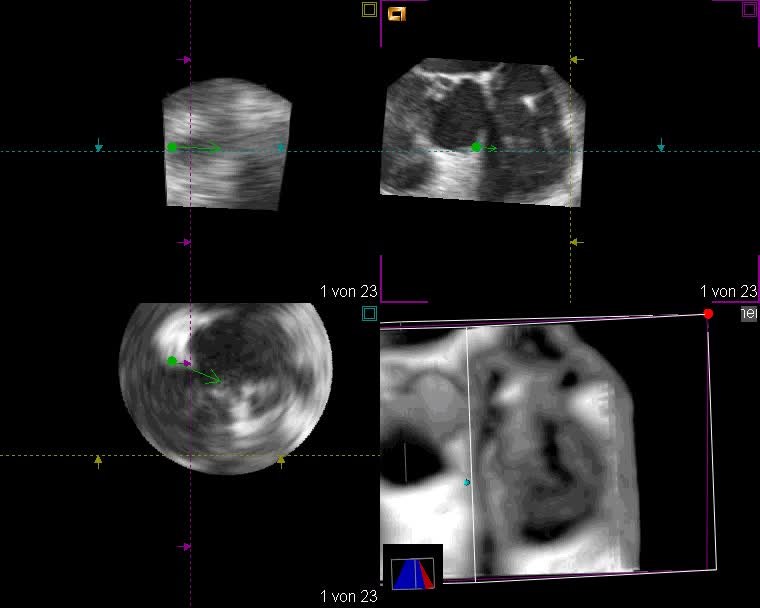
English: "The worm" presented in three-dimensional offline reconstruction of the two-dimensional transesophageal data. Three-dimensional offline reconstruction of the two-dimensional transesophageal data helped us to identify the origin of the thrombus. 3D visualization of the thrombus demonstrated that the mass was not attached to the leads in the RA and RV but trapped between them. |
||
| Date | |||
| Source | Maagh P, Butz T, Ziegler A, Meissner A, Prull M, Trappe H (2010). "The first three-dimensional visualization of a thrombus in transit trapped between the leads of a permanent dual-chamber pacemaker: a case report". Journal of Medical Case Reports. DOI:10.1186/1752-1947-4-359. PMID 21070629. PMC: 2994880. | ||
| Author | Maagh P, Butz T, Ziegler A, Meissner A, Prull M, Trappe H | ||
| Permission (Reusing this file) |
This file is licensed under the Creative Commons Attribution 2.0 Generic license.
|
||
| Provenance |
|
File history
Click on a date/time to view the file as it appeared at that time.
| Date/Time | Thumbnail | Dimensions | User | Comment | |
|---|---|---|---|---|---|
| current | 01:42, 20 November 2012 | 1.8 s, 760 × 608 (434 KB) | Open Access Media Importer Bot (talk | contribs) | Automatically uploaded media file from Open Access source. Please report problems or suggestions here. |
You cannot overwrite this file.
File usage on Commons
There are no pages that use this file.