File:The-neuroepithelial-basement-membrane-serves-as-a-boundary-and-a-substrate-for-neuron-migration-in-1749-8104-5-9-S4.ogv
Size of this JPG preview of this OGG file: 800 × 533 pixels. Other resolutions: 320 × 213 pixels | 640 × 426 pixels | 1,081 × 720 pixels.
Original file (Ogg Theora video file, length 6.5 s, 1,081 × 720 pixels, 383 kbps, file size: 304 KB)
Captions
Captions
Add a one-line explanation of what this file represents
Summary
edit| Description |
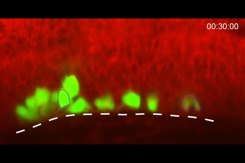
English: Timelapse movie of wild-type tg(isl1:GFP) embryo counterstained with BODIPY TR methyl ester dye. The movie was started at 24 hpf. Images were taken every 5 minutes. The movie runs at 2 frames per second. The embryo is shown in lateral view. White line delineates the ventral limit of the hindbrain. The blue outlined cell moves ventrally while the magenta outlined cell moves posteriorly. |
||
| Date | |||
| Source | Grant P, Moens C (2010). "The neuroepithelial basement membrane serves as a boundary and a substrate for neuron migration in the zebrafish hindbrain". Neural Development. DOI:10.1186/1749-8104-5-9. PMID 20350296. PMC: 2857861. | ||
| Author | Grant P, Moens C | ||
| Permission (Reusing this file) |
This file is licensed under the Creative Commons Attribution 2.0 Generic license.
|
||
| Provenance |
|
File history
Click on a date/time to view the file as it appeared at that time.
| Date/Time | Thumbnail | Dimensions | User | Comment | |
|---|---|---|---|---|---|
| current | 03:31, 20 November 2012 | 6.5 s, 1,081 × 720 (304 KB) | Open Access Media Importer Bot (talk | contribs) | Automatically uploaded media file from Open Access source. Please report problems or suggestions here. |
You cannot overwrite this file.
File usage on Commons
There are no pages that use this file.