File:Three-Dimensional-Neuroepithelial-Culture-from-Human-Embryonic-Stem-Cells-and-Its-Use-for-pone.0054552.s008.ogv
Size of this JPG preview of this OGG file: 600 × 600 pixels. Other resolutions: 240 × 240 pixels | 480 × 480 pixels | 768 × 768 pixels | 1,024 × 1,024 pixels.
Original file (Ogg Theora video file, length 7.5 s, 1,024 × 1,024 pixels, 1.33 Mbps, file size: 1.19 MB)
Captions
Captions
Add a one-line explanation of what this file represents
Summary
edit| Description |
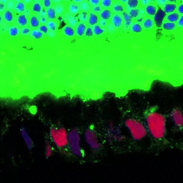
English: A movie of serial confocal images through z-axis showing GFP-labeled outer segments (green) inside hESC-derived pigmented cells in the retinal explant co-culture model. Each image of the z-stack was merged of RHODOPSIN-GFP (green), human nuclei (red), and Hoechst (blue) images. |
||
| Date | |||
| Source | Movie S2 from Zhu Y, Carido M, Meinhardt A, Kurth T, Karl M, Ader M, Tanaka E (2013). "Three-Dimensional Neuroepithelial Culture from Human Embryonic Stem Cells and Its Use for Quantitative Conversion to Retinal Pigment Epithelium". PLOS ONE. DOI:10.1371/journal.pone.0054552. PMC: 3554725. | ||
| Author | Zhu Y, Carido M, Meinhardt A, Kurth T, Karl M, Ader M, Tanaka E | ||
| Permission (Reusing this file) |
|
||
| Provenance |
|
File history
Click on a date/time to view the file as it appeared at that time.
| Date/Time | Thumbnail | Dimensions | User | Comment | |
|---|---|---|---|---|---|
| current | 19:06, 29 January 2013 | 7.5 s, 1,024 × 1,024 (1.19 MB) | Open Access Media Importer Bot (talk | contribs) | Automatically uploaded media file from Open Access source. Please report problems or suggestions here. |
You cannot overwrite this file.
File usage on Commons
There are no pages that use this file.