Category:Embryonic development
process by which the embryo is formed and develops | |||||
| Upload media | |||||
| Subclass of | |||||
|---|---|---|---|---|---|
| Has part(s) | |||||
| |||||
Subcategories
This category has the following 10 subcategories, out of 10 total.
A
- Animal pole (8 F)
B
- Blastomeres (21 F)
D
L
- Limb buds (10 F)
M
- Microspore embryogenesis (6 F)
N
- Notochord (80 F)
O
V
- Vegetal pole (12 F)
Media in category "Embryonic development"
The following 200 files are in this category, out of 280 total.
(previous page) (next page)-
 1-day-old newt egg.jpg 603 × 396; 86 KB
-
 10-day-old newt eg.jpg 839 × 547; 40 KB
-
 12-day-old newt egg.jpg 983 × 716; 50 KB
-
 14-day-old newt egg.jpg 800 × 534; 40 KB
-
 15-day-old newt egg.jpg 800 × 441; 13 KB
-
 2-day-old newt egg.jpg 800 × 575; 34 KB
-
 20-day-old newt larva.jpg 800 × 587; 41 KB
-
 2904 Preembryonic Development-02.jpg 1,957 × 1,792; 1.12 MB
-
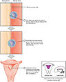 2905 Implantation.jpg 1,950 × 2,440; 1.04 MB
-
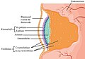 2907 Embroyonic Disc, Amniotic Cavity, Yolk Sac-02-NLtxt.jpg 1,503 × 1,055; 689 KB
-
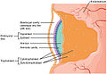 2907 Embroyonic Disc, Amniotic Cavity, Yolk Sac-02.jpg 1,529 × 1,055; 551 KB
-
 2908 Germ Layers-02-nltxt.jpg 1,854 × 1,658; 1.45 MB
-
 2908 Germ Layers-02.jpg 1,781 × 1,590; 1.06 MB
-
 2909 Embryo Week 3-02 NLtxt.jpg 1,446 × 1,276; 855 KB
-
 2909 Embryo Week 3-02.jpg 1,447 × 1,252; 634 KB
-
 2913 Embryonic Folding.jpg 1,894 × 1,735; 831 KB
-
 3-day-old newt egg.jpg 800 × 523; 32 KB
-
 3311.fig.1.jpg 1,170 × 612; 218 KB
-
 5-day-old newt egg.jpg 731 × 532; 35 KB
-
 6-day-old newt egg.jpg 800 × 619; 41 KB
-
 8-day-old newt egg.jpg 800 × 571; 34 KB
-

-
 A human embryo of 2 mm. in median sagittal section.jpg 838 × 1,006; 310 KB
-

-

-
-
-
A-Novel-Role-for-MAPKAPK2-in-Morphogenesis-during-Zebrafish-Development-pgen.1000413.s001.ogv 7.3 s, 900 × 470; 1.97 MB
-
A-Novel-Role-for-MAPKAPK2-in-Morphogenesis-during-Zebrafish-Development-pgen.1000413.s002.ogv 3.7 s, 432 × 208; 74 KB
-
A-Novel-Role-for-MAPKAPK2-in-Morphogenesis-during-Zebrafish-Development-pgen.1000413.s003.ogv 2.8 s, 432 × 208; 44 KB
-
A-Novel-Role-for-MAPKAPK2-in-Morphogenesis-during-Zebrafish-Development-pgen.1000413.s004.ogv 10 s, 658 × 517; 3.91 MB
-
A-Novel-Role-for-MAPKAPK2-in-Morphogenesis-during-Zebrafish-Development-pgen.1000413.s005.ogv 8.5 s, 600 × 300; 7.4 MB
-
A-Novel-Role-for-MAPKAPK2-in-Morphogenesis-during-Zebrafish-Development-pgen.1000413.s006.ogv 19 s, 400 × 300; 135 KB
-
 Abatus cordatus Developmental stages.jpg 1,311 × 1,679; 1.1 MB
-
 Absence of the portal system in a first trimester human.jpg 3,048 × 1,840; 2.58 MB
-
Acute-Drug-Treatment-in-the-Early-C.-elegans-Embryo-pone.0024656.s004.ogv 5.6 s, 640 × 208; 21 KB
-
Acute-Drug-Treatment-in-the-Early-C.-elegans-Embryo-pone.0024656.s005.ogv 2.2 s, 350 × 210; 14 KB
-
Acute-Drug-Treatment-in-the-Early-C.-elegans-Embryo-pone.0024656.s006.ogv 7.2 s, 350 × 210; 17 KB
-
Acute-Drug-Treatment-in-the-Early-C.-elegans-Embryo-pone.0024656.s007.ogv 7.6 s, 640 × 202; 50 KB
-
Acute-Drug-Treatment-in-the-Early-C.-elegans-Embryo-pone.0024656.s008.ogv 0.7 s, 640 × 208; 17 KB
-
Acute-Drug-Treatment-in-the-Early-C.-elegans-Embryo-pone.0024656.s009.ogv 1.2 s, 640 × 208; 17 KB
-
 Agenesis of ductus venosus human.jpg 3,240 × 1,000; 2.1 MB
-
-

-
-
-
-
-
-
-
-
-
An-automated-microfluidic-platform-for-C.-elegans-embryo-arraying-phenotypingand-long-term-live-srep10192-s5.ogv 29 s, 1,344 × 1,024; 51.2 MB
-
An-automated-microfluidic-platform-for-C.-elegans-embryo-arraying-phenotypingand-long-term-live-srep10192-s6.ogv 1 min 2 s, 658 × 524; 31.81 MB
-
An-automated-microfluidic-platform-for-C.-elegans-embryo-arraying-phenotypingand-long-term-live-srep10192-s7.ogv 40 s, 2,100 × 1,361; 12.4 MB
-
-
-
-
 Anatomic and histopathological aspects of FT organs human.jpg 3,948 × 1,292; 4.99 MB
-
 Arquénteron.jpg 1,066 × 1,061; 73 KB
-
-
-

-

-
-
-
-

-

-

-
-

-

-
 Cent uw ściana z embriogenezą.jpg 4,112 × 3,024; 4.66 MB
-
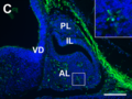
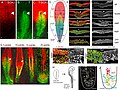 Characterization of the SOX2T-positive territory of the epiblast in chicken embryo.jpg 2,113 × 1,581; 1.21 MB
-
Collective-Motion-of-Cells-Mediates-Segregation-and-Pattern-Formation-in-Co-Cultures-pone.0031711.s001.ogv 25 s, 1,024 × 768; 23.14 MB
-
Collective-Motion-of-Cells-Mediates-Segregation-and-Pattern-Formation-in-Co-Cultures-pone.0031711.s002.ogv 12 s, 1,024 × 768; 14.4 MB
-
-
Collective-Motion-of-Cells-Mediates-Segregation-and-Pattern-Formation-in-Co-Cultures-pone.0031711.s004.ogv 0.0 s, 512 × 340; 13.15 MB
-
Collective-Motion-of-Cells-Mediates-Segregation-and-Pattern-Formation-in-Co-Cultures-pone.0031711.s005.ogv 8.1 s, 1,024 × 768; 43.09 MB
-
Collective-Motion-of-Cells-Mediates-Segregation-and-Pattern-Formation-in-Co-Cultures-pone.0031711.s006.ogv 8.0 s, 1,384 × 1,040; 48.61 MB
-
Collective-Motion-of-Cells-Mediates-Segregation-and-Pattern-Formation-in-Co-Cultures-pone.0031711.s007.ogv 15 s, 1,024 × 768; 53.72 MB
-
Collective-Motion-of-Cells-Mediates-Segregation-and-Pattern-Formation-in-Co-Cultures-pone.0031711.s008.ogv 11 s, 1,024 × 768; 58.71 MB
-
-

-

-
 Cortical neurogenesis in the mouse embryo.png 1,143 × 815; 530 KB
-
 CpG methylation in mouse development.png 1,660 × 807; 188 KB
-
-
 Developing placenta.jpg 678 × 334; 81 KB
-
 Diagrams showing the development of the amnion, chorion and allantois.jpg 1,269 × 1,151; 605 KB
-
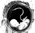
 Die Gartenlaube (1878) b 528.jpg 1,244 × 2,143; 802 KB
-
 Different degrees of EMT correlate with different tissue morphologies a.jpg 968 × 1,292; 576 KB
-
 Diversity of vertebrate gastrulation.jpg 1,073 × 1,021; 415 KB
-
 Dose-dependent reshaping of primitive streak.jpg 968 × 1,236; 743 KB
-
-
Drosophila cleavage and gastrulation.webm 30 s, 1,920 × 800; 42.28 MB
-
Dynamics of mesodermal cell ingression chicken embryo.ogv 12 s, 226 × 720; 4.2 MB
-
 Early development stages with names.gif 311 × 360; 3.2 MB
-
 Early gastrulation in amphibian embryos.png 3,570 × 1,358; 1,007 KB
-
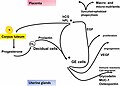 Early hormonal interaction after implantation.jpg 1,663 × 1,189; 125 KB
-

-
Echinoderm development.webm 3 min 3 s, 640 × 480; 38.31 MB
-
 Echinoderm embryo undergoing second cleavage.jpg 1,740 × 1,700; 532 KB
-
 Embryo 2 (PSF).png 3,551 × 2,027; 319 KB
-
 Embryogenesis-es2.svg 512 × 384; 324 KB
-
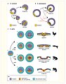
 Embryogenesis.jpg 2,424 × 2,859; 3.27 MB
-
 Embryogenesis1.jpg 1,220 × 1,430; 359 KB
-
 Embryological development of the human venous system.png 2,980 × 1,672; 1.37 MB
-
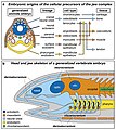
 Embryonic and pelagic stages of select neuston.jpg 1,498 × 1,015; 354 KB
-
Embryonic development of a salamander, filmed in the 1920s.ogv 14 min 52 s, 400 × 300; 59.31 MB
-
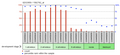 EmbyronicDevelopmentMicroarray.png 1,328 × 614; 102 KB
-
-
-
-
-
-

-
 Experimental manipulation of the gastrulation mode in different organisms.jpg 1,166 × 1,480; 850 KB
-

-
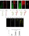
 Expression patterns of L. fluviatilis NogginA (a–j), NogginC (k–r) and NogginD (s–v).jpg 1,495 × 2,037; 1.69 MB
-
-
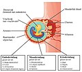
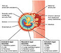
 Fate of germ layers of the embryo.png 500 × 433; 183 KB
-
 Formation and patterning of the mouse neural tube.png 1,305 × 1,505; 636 KB
-
 Formation of the primitive body plan following gastrulation in the mouse.png 1,279 × 1,187; 1,016 KB
-
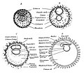 Four diagrams showing hypothetical stages of early human embryos.jpg 1,631 × 1,434; 943 KB
-
-
-
-
-
-
-
-
-
 Gastrulation forms in vertebrates.jpeg 1,280 × 1,173; 88 KB
-

-
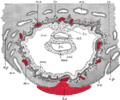 Gray32.png 500 × 417; 53 KB
-
 Hans Spemann.jpg 849 × 1,371; 500 KB
-
-
-
-
-
-
 Hipófisis embrionária e14,5.png 879 × 657; 709 KB
-
 Histological appearance of the FT liver with normal PVS human.jpg 1,978 × 1,078; 2.72 MB
-
 Histopathological and ultrasound aspect of normal FT liver human.jpg 4,016 × 1,092; 1.95 MB
-
 How the Turtle Gets its Shell.svg 2,122 × 1,568; 54 KB
-

-
 Human embryo Section of embryonic rudiment in Peters' ovum (first week).jpg 1,141 × 857; 540 KB
-

-

-

-
-
-

-
 Implanting embryo.jpg 1,497 × 2,381; 215 KB
-
Invasiveness-of-mouse-embryos-to-human-ovarian-cancer-cells-HO8910PM-and-the-role-of-MMP-9-1475-2867-12-23-S1.ogv 1 min 24 s, 375 × 288; 22.21 MB
-
-
-
Late embryonic development in Caenorhabditis elegans - pbio.1001115.s012.ogv 6.3 s, 516 × 486; 1.19 MB
-
Latrunculin A affects actin cytoskeleton in the whole embryonic disc (01).ogv 5.8 s, 694 × 520; 2.23 MB
-
 Latrunculin A affects actin cytoskeleton in the whole embryonic disc.jpeg 1,280 × 727; 159 KB
-
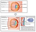
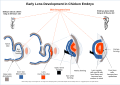 Lens embryogenisis.svg 1,488 × 1,052; 242 KB
-

-
Long-term tracking of a nuclear red stained chicken embryo.ogv 10 s, 248 × 718; 4.63 MB
-
Long-term tracking of the mesodermal progenitors chicken embryo.ogv 10 s, 236 × 432; 889 KB
-
-
Longevity of tracks along the primitive streak (PS) chicken embryo.ogv 5.9 s, 286 × 720; 1.48 MB
-
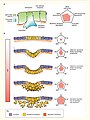
 Mesoderm-ring to -crescent transition.jpg 1,173 × 1,826; 1.01 MB
-
 Meyers b5 s0349 b1.png 486 × 234; 28 KB
-
 Meyers b5 s0682 b1.png 474 × 789; 304 KB
-
 Migration of Neural Crest Cells (v2).jpg 3,024 × 2,297; 867 KB
-
-
-
-

-
Neurergus kaiseri.webm 10 s, 960 × 540; 182 KB
-

-

-
Novel-PRD-like-homeodomain-transcription-factors-and-retrotransposon-elements-in-early-human-ncomms9207-s8.ogv 2 min 3 s, 640 × 480; 17.36 MB
-
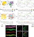
 Number of TSOX2 double-positive cells during early chicken stages embryo.jpg 2,007 × 2,502; 828 KB
-
-

-
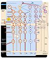
 Outline of key signaling pathways.png 4,148 × 1,682; 1.3 MB
-
 Parathyroid glands during embryogenesis.jpg 1,000 × 495; 63 KB
-
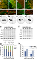
 Phalangium opilio labial gene expression.jpg 4,000 × 2,519; 2.65 MB
-
 Pharyngal arches.gif 314 × 379; 1.54 MB
-
-
 Placenta formation.jpg 339 × 561; 51 KB
-
 Platelets in cancer EMT.jpg 1,280 × 720; 96 KB
-
 Portal venous system human.jpg 3,940 × 1,192; 3 MB
-

-
Pre-gastrulation cell movements are impaired by ROCK inhibition (01).ogv 10 s, 1,041 × 780; 7.31 MB
-
Pre-gastrulation cell movements are impaired by ROCK inhibition (02).ogv 9.5 s, 1,041 × 780; 6.62 MB
-
 Pre-gastrulation-cell-movements-are-impaired-by-ROCK-inhibition-A-D-F-I-Dorsal-views a.jpg 1,233 × 1,293; 1.14 MB
-
-

-
 Primitiv Node.jpg 720 × 504; 42 KB
-
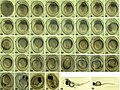 Pseudorasbora parva (10.3897-zoologia.35.e22162) Figures 2–39.jpg 1,997 × 1,494; 1.42 MB
-
 PSM V71 D516 A primitive muscular segment of a cat embryo.png 503 × 715; 161 KB


_epiblast_chicken_embryo.jpg)


_b_528.jpg)


_epithelial_phenotype_during_development_chicken_embryo.jpg)


.png)


.png)


_signature_genes_in_chicken_and_mouse_embryos.jpg)
,_NogginC_(k%E2%80%93r)_and_NogginD_(s%E2%80%93v).jpg)


.jpg)


_from_Ornament_to_the_Mind_of_Medicine_Buddha-_Blue_Beryl_Lamp_Illuminating_Four_Tantras_written_around_the_year_1720_by_Desi_Sangye_Gyatso_(1653%E2%80%931705),_the_regent_(Desi)_of_the_5th_Dalai_Lama.png)


.jpg)


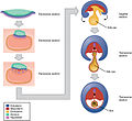
_The_lower_panel_shows_the_Hensen%E2%80%99s_node_(Chick_embryo),_embryonic_shield_(Fish_embryo),_and_nodeAVE_(Mouse_embryo)..png)


_progenitor_territories_results_in_NMPs_remaining_as_the_major_PS_remnant_in_the_tail_bud.jpg)


_Figures_2%E2%80%9339.jpg)
