Category:Fibroblasts
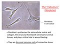
most common cell of connective tissue in animal, that synthesizes the extracellular matrix and collagen, the structural framework (stroma) for animal tissues, and plays a critical role in wound healing  | |||||
| Upload media | |||||
| Instance of | |||||
|---|---|---|---|---|---|
| Subclass of |
| ||||
| |||||
Subcategories
This category has the following 5 subcategories, out of 5 total.
Media in category "Fibroblasts"
The following 181 files are in this category, out of 181 total.
-
 1 Alien Planet Surface.tif 1,388 × 1,040; 4.14 MB
-
-
-
-
-
-
 Actin filaments of cardiac fibroblasts.jpg 3,456 × 4,608; 7.35 MB
-
-
-
Adaptive-Stress-Response-in-Segmental-Progeria-Resembles-Long-Lived-Dwarfism-and-Calorie-pgen.0020192.sv001.ogv 1 min 54 s, 240 × 180; 4.9 MB
-
-
An-ES-Like-Pluripotent-State-in-FGF-Dependent-Murine-iPS-cells-pone.0016092.s006.ogv 9.9 s, 312 × 240; 1.06 MB
-
Arp23-complex-activity-in-filopodia-of-spreading-cells-1471-2121-9-65-S1.ogv 1 min 5 s, 434 × 440; 1.65 MB
-
Arp23-complex-activity-in-filopodia-of-spreading-cells-1471-2121-9-65-S4.ogv 3.8 s, 210 × 288; 152 KB
-
Arp23-complex-activity-in-filopodia-of-spreading-cells-1471-2121-9-65-S5.ogv 20 s, 750 × 666; 418 KB
-
Arp23-complex-activity-in-filopodia-of-spreading-cells-1471-2121-9-65-S6.ogv 21 s, 614 × 394; 2.49 MB
-
-
-
-
BH3-Peptides-Induce-Mitochondrial-Fission-and-Cell-Death-Independent-of-BAXBAK-pone.0005646.s005.ogv 5.0 s, 370 × 411; 112 KB
-
-
-
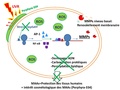
 Cancer-associated Fibroblasts.png 1,026 × 1,060; 195 KB
-
 Cell expantion.jpg 2,452 × 1,756; 6.21 MB
-

-
-
-
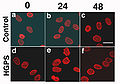
 Confocal analysis of dermal fibroblasts after heat shock stress (progeria).jpg 900 × 1,347; 273 KB
-
-
-
-
-
 Culture de fibroblastes humains.tif 15,937 × 15,730; 230.62 MB
-
-
-
-
-
-
Development-of-a-macromolecular-prodrug-for-the-treatment-of-inflammatory-arthritis-mechanisms-ar3130-S1.ogv 8.3 s, 992 × 1,040; 689 KB
-
 Diferenciación de fibroblastos a miofibroblastos cardíacos.jpg 10,618 × 2,336; 8.88 MB
-
Direct-Generation-of-Neurosphere-Like-Cells-from-Human-Dermal-Fibroblasts-pone.0021801.s010.ogv 56 s, 640 × 480; 9.47 MB
-
-
-
-
-
 Double Star Satellite FLU.tif 1,388 × 1,040; 1.13 MB
-
 Effet de Porphyra-334 sur les fibroblastes humaines exposés aux rayons UVA.pdf 1,500 × 1,125; 260 KB
-
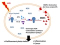 Effet du porphyra-334 sur des fibroblastes humains exposés aux UVA.pdf 1,500 × 1,125, 2 pages; 285 KB
-
-
-
-
 Fibroblast (BPAE).jpg 3,072 × 2,048; 2.79 MB
-
 Fibroblast cells.jpg 691 × 516; 79 KB
-
 Fibroblast focus.jpg 2,040 × 1,536; 2.08 MB
-
 Fibroblast,polydopamine nanoparticles and mithocondria.jpg 2,048 × 2,048; 579 KB
-
 Fibroblast-2.jpg 2,999 × 2,249; 693 KB
-
 Fibroblastid (BPAE).jpg 3,304 × 2,202; 2.73 MB
-
 Fibroblastid.jpg 6,000 × 4,627; 7.53 MB
-
 Fibroblastos 100x a.jpg 2,048 × 1,536; 257 KB
-
 Fibroblastos 100x b.jpg 2,048 × 1,536; 244 KB
-
 Fluorescent image fibroblast.jpg 1,500 × 2,250; 440 KB
-
 Focaladhesion.jpg 428 × 441; 90 KB
-
 Formation of the new blood vessels 2.jpg 1,920 × 1,080; 507 KB
-
 Formation of the new blood vessels.jpg 1,920 × 1,200; 2.81 MB
-
 Galaxy and Black Hole.tif 1,024 × 1,024; 3 MB
-
-
-
-
-
 Human Cell Groups distributed by Cell Count and by Aggregate Cell Mass.jpg 3,162 × 2,096; 1.08 MB
-
 Human Cell Groups; Cell Count, Cell Mass, and Aggregate Cell Mass (Biomass).png 1,258 × 847; 149 KB
-
-
-

-
 Humanstemcell.JPG 3,072 × 2,304; 2.41 MB
-
 Ice Planet Treasures (2).tif 1,388 × 1,040; 4.14 MB
-
 Indian Muntjac fibroblast cells (23725924864).jpg 1,924 × 1,218; 718 KB
-
-
-
-
-
-
-
-
-
-
-
-
 Macrófago o célula de Langerhans a 40x.jpg 2,048 × 1,536; 325 KB
-
-
-
-
-
-
-
 Miofibroblasto 1B MET.png 355 × 343; 171 KB
-
 Miofibroblasto1A MET.png 310 × 381; 259 KB
-
Movies-of-cellular-and-sub-cellular-motion-by-digital-holographic-microscopy-1475-925X-5-21-S1.ogv 3.5 s, 432 × 432; 1.24 MB
-
Movies-of-cellular-and-sub-cellular-motion-by-digital-holographic-microscopy-1475-925X-5-21-S2.ogv 6.3 s, 464 × 464; 2.75 MB
-
Movies-of-cellular-and-sub-cellular-motion-by-digital-holographic-microscopy-1475-925X-5-21-S3.ogv 6.3 s, 464 × 464; 1.24 MB
-
Movies-of-cellular-and-sub-cellular-motion-by-digital-holographic-microscopy-1475-925X-5-21-S4.ogv 6.3 s, 464 × 464; 1.59 MB
-
Movies-of-cellular-and-sub-cellular-motion-by-digital-holographic-microscopy-1475-925X-5-21-S5.ogv 6.3 s, 452 × 452; 2.94 MB
-
Movies-of-cellular-and-sub-cellular-motion-by-digital-holographic-microscopy-1475-925X-5-21-S6.ogv 6.3 s, 452 × 452; 1.44 MB
-
Movies-of-cellular-and-sub-cellular-motion-by-digital-holographic-microscopy-1475-925X-5-21-S7.ogv 6.5 s, 452 × 452; 2.17 MB
-
-

-
-
-
 Neonatal Human Dermal Fibroblasts.jpg 1,256 × 949; 129 KB
-
 Network of Life FLU.tif 1,388 × 1,040; 4.13 MB
-
 Nucleus of cardiac fibroblasts.jpg 3,456 × 4,608; 4.44 MB
-
 Oct4KOT1DAPIT2Acta2T3Oct4T4eYFP1 500IgGOct4IgGeYFP 3.tif 512 × 512; 769 KB
-
-
-
-
-
-
-
Phosphorylation-of-p130Cas-initiates-Rac-activation-and-membrane-ruffling-1471-2121-9-50-S1.ogv 2.1 s, 736 × 768; 416 KB
-
Phosphorylation-of-p130Cas-initiates-Rac-activation-and-membrane-ruffling-1471-2121-9-50-S2.ogv 2.1 s, 1,264 × 1,008; 1.36 MB
-
Phosphorylation-of-p130Cas-initiates-Rac-activation-and-membrane-ruffling-1471-2121-9-50-S3.ogv 2.1 s, 848 × 896; 776 KB
-
Phosphorylation-of-p130Cas-initiates-Rac-activation-and-membrane-ruffling-1471-2121-9-50-S4.ogv 2.1 s, 848 × 896; 948 KB
-
Phosphorylation-of-p130Cas-initiates-Rac-activation-and-membrane-ruffling-1471-2121-9-50-S5.ogv 2.1 s, 848 × 896; 480 KB
-
Physically-Induced-Cytoskeleton-Remodeling-of-Cells-in-Three-Dimensional-Culture-pone.0045512.s014.ogv 8.0 s, 1,024 × 1,024; 877 KB
-
Probing-Cellular-Dynamics-with-a-Chemical-Signal-Generator-pone.0004847.s002.ogv 26 s, 800 × 600; 495 KB
-
Probing-Cellular-Dynamics-with-a-Chemical-Signal-Generator-pone.0004847.s003.ogv 23 s, 800 × 600; 229 KB
-
-
-
-
-
-
-
-
-
-
-
-
-
-
-
 RDEBFs Transfected with Lipoplexes.tiff 1,392 × 1,040; 2.46 MB
-
-
-
-
-
-
-
-
-
-
-
-
-
-
 Test d'invasion de fibroblastes par des parasites Toxoplasma gondii.jpg 2,625 × 2,006; 549 KB
-
 The creation of Life.jpg 1,280 × 960; 713 KB
-
-
-
-
-
-
-
-
-
-
-
Theoretical-Model-for-Cellular-Shapes-Driven-by-Protrusive-and-Adhesive-Forces-pcbi.1001127.s003.ogv 4.6 s, 508 × 364; 435 KB
-
Theoretical-Model-for-Cellular-Shapes-Driven-by-Protrusive-and-Adhesive-Forces-pcbi.1001127.s004.ogv 5.3 s, 508 × 364; 228 KB
-
-
-

-
 Vimentin.jpg 2,304 × 3,072; 2.06 MB
-
Virion-Assembly-Factories-in-the-Nucleus-of-Polyomavirus-Infected-Cells-ppat.1002630.s002.ogv 54 s, 640 × 480; 24.06 MB
-
Virion-Assembly-Factories-in-the-Nucleus-of-Polyomavirus-Infected-Cells-ppat.1002630.s003.ogv 34 s, 960 × 720; 25.45 MB
-
-
-
-
-
-
 WI-38-Li-and-Tollefsbol-2011.gif 600 × 334; 113 KB
-


.jpg)
_CROPPED.jpg)
.jpg)
.jpg)


.jpg)


.png)


.jpg)


_%D1%84%D1%96%D0%B1%D1%80%D0%BE%D0%B1%D0%BB%D0%B0%D1%81%D1%82%D1%96%D0%B2_%D1%81%D0%B5%D1%80%D1%86%D1%8F_%D1%89%D1%83%D1%80%D0%B0.jpg)
.jpg)