File:Pathology and treatment of diseases of women (1912) (14758654596).jpg
Original file(1,906 × 1,840 pixels, file size: 1.05 MB, MIME type: image/jpeg)

Captions

Captions

Add a one-line explanation of what this file represents

Summary edit

Description
English:

Identifier: pathologytreatme00mart (find matches)
Title: Pathology and treatment of diseases of women
Year: 1912 (1910s)
Authors: Martin, August Eduard, 1847- Jung, Ph. (Philipp Jacob), 1870-1918
Subjects: Gynecology Gynecology
Publisher: New York : Rebman company
Contributing Library: Francis A. Countway Library of Medicine
Digitizing Sponsor: Open Knowledge Commons and Harvard Medical School

View Book Page: Book Viewer
About This Book: Catalog Entry
View All Images: All Images From Book
Click here to view book online to see this illustration in context in a browseable online version of this book.

Text Appearing Before Image:
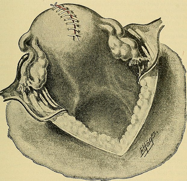
. (Abdominal operation.) mata, enucleation is executed by opening the peritoneum and the capsuleat the apex of the tumor (Fig. 120). If several myomata have formed independently of each other, thenfrom the bed of the first enucleated tumor the second myoma is incised.If the tumors are too far apart from each other separate incisions aremade at such points on the surface as appear suitable. I seize thetumors with bullet- or Muzeux forceps; the introduction of drill in-struments only retards the process of the operation. The tumor, now, is PATHOLOGY OF THE VAGINA AND UTERUS 263 pulled upward and peeled out with the fingers. Stronger bands, connect-ing the growth with the capsule of the tumor are cut with knife or scis-sors. The bed of the tumor mostly does not bleed. The hemorrhagestops, at all events, if the assistant presses the cervix with both handsfrom the sides. Otherwise use artery clamps. The bed of the tumor issmoothed as much as possible, thereby the capsule is removed in most
Text Appearing After Image:
Fig. 122.—The uterus wound is sutured. cases. Henkel196 has recommended the systematic excision of the capsule.The uterine mucosa is examined thoroughly after opening of the uterinecavity. Abrasio mucosae through the existing opening, and eventuallyresection of the superfluous parts of the mucosa. Closure of the uterinecavity by a continued submucous catgut stitch (Fig. 121). Suturing ofthe bed of the myoma with continued sutures in rows. Coaptation ofthe wound margins by a continuous catgut stitch (Fig. 122). The en-tire body of the uterus is palpated for myoma-nuclei. Diseased ovariesand tubes are removed or resected. Examination - of the, peritoneal 264 DISEASES OF WOMEN cavity for changes previously unnoticed. Replacing- of the uterus,cleansing of the abdominal cavity, suturing of the peritoneum with con-tinued suture after placing a sterile cloth on the intestines which tendto press forward, which is removed, before the last stitch through theupper end of the peritoneal wound i

Note About Images

Please note that these images are extracted from scanned page images that may have been digitally enhanced for readability - coloration and appearance of these illustrations may not perfectly resemble the original work.
Date
Source

https://www.flickr.com/photos/internetarchivebookimages/14758654596/

Author Internet Archive Book Images
Permission
(Reusing this file)
At the time of upload, the image license was automatically confirmed using the Flickr API. For more information see Flickr API detail.
Flickr tags
InfoField
  • bookid:pathologytreatme00mart
  • bookyear:1912
  • bookdecade:1910
  • bookcentury:1900
  • bookauthor:Martin__August_Eduard__1847_
  • bookauthor:Jung__Ph___Philipp_Jacob___1870_1918
  • booksubject:Gynecology
  • bookpublisher:New_York___Rebman_company
  • bookcontributor:Francis_A__Countway_Library_of_Medicine
  • booksponsor:Open_Knowledge_Commons_and_Harvard_Medical_School
  • bookleafnumber:282
  • bookcollection:medicalheritagelibrary
  • bookcollection:francisacountwaylibrary
  • bookcollection:americana
Flickr posted date
InfoField
30 July 2014


Licensing edit

This image was taken from Flickr's The Commons. The uploading organization may have various reasons for determining that no known copyright restrictions exist, such as:
  1. The copyright is in the public domain because it has expired;
  2. The copyright was injected into the public domain for other reasons, such as failure to adhere to required formalities or conditions;
  3. The institution owns the copyright but is not interested in exercising control; or
  4. The institution has legal rights sufficient to authorize others to use the work without restrictions.

More information can be found at https://flickr.com/commons/usage/.


Please add additional copyright tags to this image if more specific information about copyright status can be determined. See Commons:Licensing for more information.
This image was originally posted to Flickr by Internet Archive Book Images at https://flickr.com/photos/126377022@N07/14758654596. It was reviewed on 23 October 2015 by FlickreviewR and was confirmed to be licensed under the terms of the No known copyright restrictions.

23 October 2015

File history

Click on a date/time to view the file as it appeared at that time.

Date/TimeThumbnailDimensionsUserComment
current09:37, 23 October 2015Thumbnail for version as of 09:37, 23 October 20151,906 × 1,840 (1.05 MB) (talk | contribs)== {{int:filedesc}} == {{information |description={{en|1=<br> '''Identifier''': pathologytreatme00mart ([https://commons.wikimedia.org/w/index.php?title=Special%3ASearch&profile=default&fulltext=Search&search=insource%3A%2Fpathologytreatme00mart%2F fin...

There are no pages that use this file.